

Ultrasound of ankles in the diagnosis of complications of chikungunya fever
- PMID: 28428648
- PMCID: PMC5396995
- DOI: 10.1590/0100-3984.2016.0221
Ultrasound of ankles in the diagnosis of complications of chikungunya fever
Abstract
Objective: To describe the main ultrasound findings of chikungunya fever in the ankle.
Materials and methods: This was a cross-sectional observational study involving 52 patients referred to the Hospital Universitário Pedro Ernesto and presenting with clinical and biochemical evidence of chikungunya fever. The examinations were performed by a radiologist with more than 20 years of experience in ultrasound.
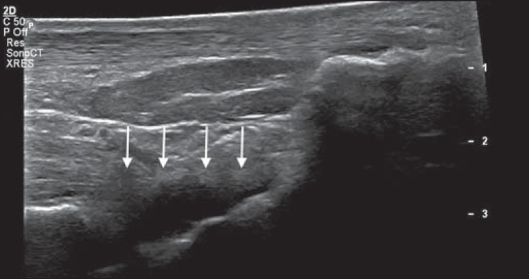
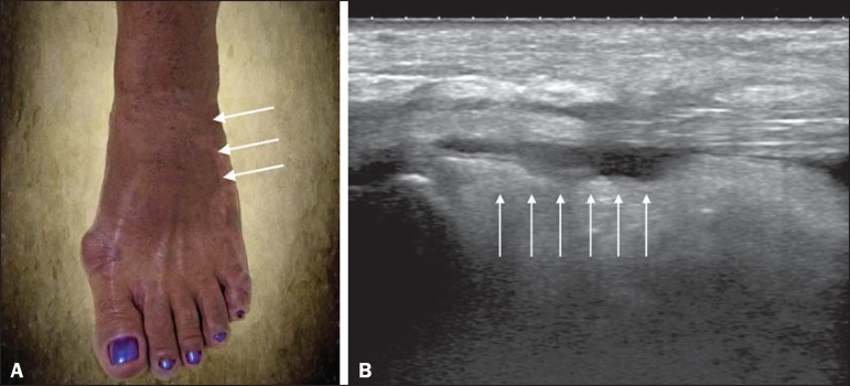
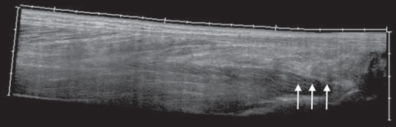
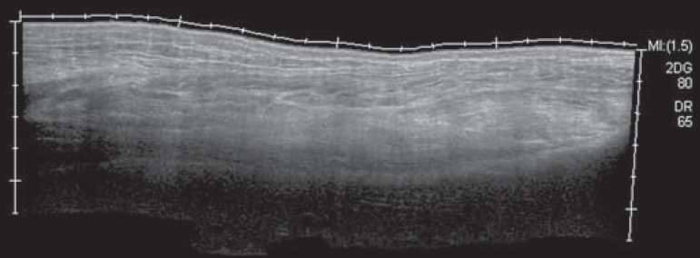
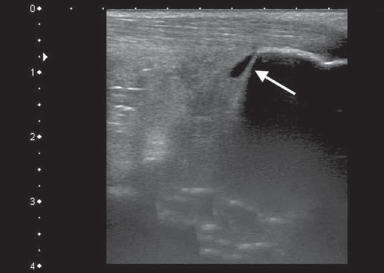
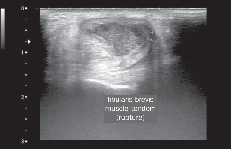
Results: The predominant gender was female (in 88.5%), and the mean age was 58.4 years. The majority (61.5%) of the patients came from the northern part of the city of Rio de Janeiro, and 46.2% were using corticosteroids to treat inflammatory symptoms. The most common alterations observed by ultrasound were joint effusion (in 69.2%), tenosynovitis (in 59.6%), cellulitis (in 46.2%), Kager's fat pad thickening (in 29.9%), myositis (of the soleus or flexor hallucis longus muscle) (in 17.3%), retrocalcaneal bursitis (in 5.8%), tendon ruptures (in 3.8%), and increased vascular flow on power Doppler (in 3.8%).
Conclusion: Signs of synovitis and tenosynovitis were the main ultrasound findings in a predominantly female population with a mean age of 58.4 years. Further studies are needed in order to define the role of ultrasound in the follow-up of such patients.
Objetivo: Descrever os principais achados ultrassonográficos da febre chikungunya no tornozelo.
Materiais e métodos: Estudo transversal e observacional com 52 pacientes encaminhados ao Hospital Universitário Pedro Ernesto com quadros clínico e laboratorial compatíveis com febre chikungunya. Os exames foram realizados por um radiologista com mais de 20 anos de experiência no método.
Resultados: Houve predomínio do sexo feminino (88,5%) e média de idade dos pacientes de 58,4 anos. A maioria dos doentes (61,5%) era proveniente da zona norte da cidade do Rio de Janeiro e fazia uso de esteroides (46,2%) para o tratamento dos sintomas inflamatórios. As alterações ultrassonográficas mais comuns foram: derrame articular (69,2%), tenossinovites (59,6%), celulite (46,2%), espessamento da gordura de Kager (29,9%), miosite (sóleo e/ou flexor longo do hálux) (17,3%), bursite retrocalcânea (5,8%), roturas tendíneas (3,8%) e hiperfluxo vascular pelo Doppler de amplitude (3,8%).
Conclusão: Predominaram os sinais ultrassonográficos de sinovite e tenossinovite numa população majoritariamente do sexo feminino e com idade média de 58,4 anos. Sugere-se a realização de outros estudos para definição do papel da ultrassonografia no acompanhamento desses doentes.
Keywords: Arboviruses; Chikungunya virus; Synovitis; Tenosynovitis; Ultrasonography.
Figures
Similar articles
-
Ultrasonography of Hands and Wrists in the Diagnosis of Complications of Chikungunya Fever.J Ultrasound Med. 2018 Feb;37(2):511-520. doi: 10.1002/jum.14344. Epub 2017 Aug 8. J Ultrasound Med. 2018. PMID: 28786505
-
The functional anatomy of Kager's fat pad in relation to retrocalcaneal problems and other hindfoot disorders.J Anat. 2006 Jan;208(1):91-7. doi: 10.1111/j.1469-7580.2006.00510.x. J Anat. 2006. PMID: 16420382 Free PMC article.
-
The Kager's fat pad radiological anatomy revised.Surg Radiol Anat. 2021 Jan;43(1):79-86. doi: 10.1007/s00276-020-02552-1. Epub 2020 Aug 19. Surg Radiol Anat. 2021. PMID: 32813031 Free PMC article.
-
Ultrasonography for diagnosis, monitoring and treatment of tenosynovitis in patients with rheumatoid arthritis.Dan Med J. 2018 Mar;65(3):B5474. Dan Med J. 2018. PMID: 29510818 Review.
-
Ultrasound and structural changes in inflammatory arthritis: synovitis and tenosynovitis.Ann N Y Acad Sci. 2009 Feb;1154:139-51. doi: 10.1111/j.1749-6632.2009.04388.x. Ann N Y Acad Sci. 2009. PMID: 19250235 Review.
Cited by
-
The Development and Evaluation of a Combined Infection-Rheumatology Assessment Service in Response to the Chikungunya Fever Epidemic.Am J Trop Med Hyg. 2023 Mar 20;108(5):1003-1006. doi: 10.4269/ajtmh.22-0698. Print 2023 May 3. Am J Trop Med Hyg. 2023. PMID: 36940667 Free PMC article.
-
Chikungunya fever and lymphedema of limbs.J Vasc Bras. 2020 Aug 7;19:e20190100. doi: 10.1590/1677-5449.190100. J Vasc Bras. 2020. PMID: 34178062 Free PMC article. No abstract available.
-
Musculoskeletal involvement in neglected tropical diseases: a comprehensive review.Skeletal Radiol. 2024 Oct;53(10):2143-2160. doi: 10.1007/s00256-024-04595-6. Epub 2024 Jan 25. Skeletal Radiol. 2024. PMID: 38267762 Review.
-
Carpal tunnel syndrome observed after an arbovirus infection: A preliminary case series report.eNeurologicalSci. 2018 Aug 21;12:31-33. doi: 10.1016/j.ensci.2018.08.004. eCollection 2018 Sep. eNeurologicalSci. 2018. PMID: 30211326 Free PMC article.
-
Disseminated intramuscular cysticercosis diagnosed incidentally in a patient with joint pain.Radiol Bras. 2019 Sep-Oct;52(5):345-346. doi: 10.1590/0100-3984.2017.0219. Radiol Bras. 2019. PMID: 31656357 Free PMC article. No abstract available.
References
-
- Imai K, Nakayama E, Maeda T, et al. Chikungunya fever in Japan imported from the Caribbean Islands. Jpn J Infect Dis. 2016;69:151–153. - PubMed
-
- Horcada ML, Díaz-Calderón C, Garrido L. Chikungunya fever. Rheumatic manifestations of an emerging disease in Europe. Reumatol Clin. 2015;11:161–164. - PubMed
-
- Weaver SC, Lecuit M. Chikungunya virus and the global spread of a mosquito-borne disease. N Engl J Med. 2015;372:1231–1239. - PubMed
-
- Javelle E, Gautret P, Simon F. Chikungunya, the emerging migratory rheumatism. Lancet Infect Dis. 2015;15:509–510. - PubMed
LinkOut - more resources
Full Text Sources
Other Literature Sources