. 2017 Mar-Apr;50(2):126-131.
doi: 10.1590/0100-3984.2015.0227.

What radiologists should know about tomographic evaluation of acute diverticulitis of the colon

Affiliations

What radiologists should know about tomographic evaluation of acute diverticulitis of the colon

Aline de Araújo Naves et al. Radiol Bras. 2017 Mar-Apr.

Abstract

Acute diverticulitis of the colon is a common indication for computed tomography, and its diagnosis and complications are essential to determining the proper treatment and establishing the prognosis. The adaptation of the surgical classification for computed tomography has allowed the extent of intestinal inflammation to be established, the computed tomography findings correlating with the indication for treatment. In addition, computed tomography has proven able to distinguish among the main differential diagnoses of diverticulitis. This pictorial essay aims to present the computed tomography technique, main radiological signs, major complications, and differential diagnoses, as well as to review the classification of acute diverticulitis.

A diverticulite aguda dos cólons é uma indicação frequente de exame tomográfico, sendo o seu diagnóstico e das suas complicações fundamental para determinar uma adequada conduta terapêutica e estabelecer o prognóstico. A adaptação da classificação cirúrgica para a tomografia computadorizada permitiu estabelecer a extensão do processo inflamatório intestinal, correlacionando o quadro tomográfico com a indicação de tratamento. Além disto, a tomografia computadorizada tem demonstrado ser capaz de distinguir os principais diagnósticos diferenciais da diverticulite aguda dos cólons. Este ensaio iconográfico tem por objetivo apresentar a técnica de exame tomográfico, os principais sinais radiológicos, e revisar a classificação e as principias complicações e diagnósticos diferenciais da diverticulite aguda dos cólons.

Keywords: Abdomen, acute; Diverticulitis, colonic; Tomography, X-ray computed.


Figures

Figure 1
Figure 1
Inflamed diverticulum. Intravenous contrast-enhanced axial CT of the abdomen, showing the diverticulum with discrete wall thickening (arrow) and increased attenuation of pericolonic fat.
Figure 2
Figure 2
Wall thickening. Intravenous and rectal contrast-enhanced coronal CT scan of the abdomen, showing colonic diverticula associated with thickening of the intestinal wall to > 1.0 cm, with an extent of 8.0 cm (arrow).
Figure 3
Figure 3
Inflammatory signs in pericolonic fat. Intravenous and rectal contrastenhanced axillary CT, in the axial plane, showing increased mesenteric fat attenuation (arrow) adjacent to the inflammatory process in the diverticula.
Figure 4
Figure 4
A,B: Rectal contrast-enhanced CT of abdomen showing signs of acute diverticulitis, characterized by sigmoid wall thickening, diverticula, and increased regional fat density (arrow in A), as well as extraluminal gas, indicating pneumoperitoneum (arrows in B). C: Signs of intestinal perforation. Intravenous contrast-enhanced abdominal CT, in the axial plane, showing pneumoretroperitoneum (arrows) secondary to diverticulitis.
Figure 5
Figure 5
Pericolonic/distal abscess. Intravenous contrast-enhanced abdominal CT, in the axial plane, showing heterogenous fluid collections (arrows) surrounded by a hyperintense halo with contrast enhancement.
Figure 6
Figure 6
Fistulas to adjacent organs. Rectal contrast-enhanced CT of the abdomen, in the sagittal plane, showing a fistulous pathway between the inflamed colonic segment and the vagina (colovaginal fistula). The diagnostic hypothesis of fistula can be suggested when there is inflammatory tissue or obliteration and increased density of the fat between the colon and the adjacent organs, as well as intraluminal gas (in the bladder, vaginal canal, or other lumen).
Figure 7
Figure 7
Hinchey stage 0. Intravenous contrast-enhanced abdominal CT, in the axial plane, showing colonic diverticula (arrow), with discrete wall thickening.
Figure 8
Figure 8
Hinchey stage Ia. Abdominal CT, in the coronal plane, without contrast. Note the wall thickening of the descending colon, accompanied by a perforated diverticulum at the mesenteric border (arrow) and increased density of the adjacent fat, without any fluid collections.
Figure 9
Figure 9
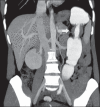
Hinchey stage Ib. Intravenous contrast-enhanced abdominal CT, in the coronal plane, showing wall thickening of the sigmoid, with an adjacent pericolonic abscess (arrows).
Figure 10
Figure 10
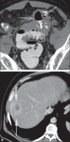
Hinchey stage II. Intravenous and rectal contrast-enhanced axial CT of the abdomen. Note the thickened and finely heterogeneous walls of the sigmoid (arrows in A) and the hepatic abscess (arrows in B) related to the inflammatory process in the colon.
Figure 11
Figure 11
Hinchey stage III. Intravenous and rectal contrast-enhanced axial CT of the abdomen showing diverticulitis with multiple abscesses (arrow) in the inframesocolic region and pneumoperitoneum, together with generalized peritonitis.
Figure 12
Figure 12
Intravenous contrast-enhanced axial CT of the abdomen showing diverticulitis with perforation and pelvic abscesses with heterogeneous content (arrow) in a patient who subsequently underwent surgery, during which a large amount of pus and feces were found in the peritoneal cavity.
Figure 13
Figure 13
Thrombosis of the portal vein. Intravenous and rectal contrast-enhanced axial CT of the abdomen showing a thrombus within the superior mesenteric vein (arrow in A), together with acute perforated and blocked diverticulitis (arrow in B), in a patient with diabetes.
Figure 14
Figure 14
Adenocarcinoma of the colon. Intravenous and rectal contrast-enhanced coronal CT of the abdomen, showing asymmetric wall thickening in the descending colon (arrow), with an abrupt transition to the normal loop (the shoulder sign)

References

    1. Tiferes DA, Jayanthi SK, Liguori AAL. D'Ippolito G, Caldana RP. Gastrointestinal. 1ª ed. Rio de Janeiro: Editora Sarvier; 2011. Cólon, reto e apêndice; pp. 203–252. Série CBR.
    1. Andeweg CS, Mulder IM, Felt-Bersma RJ, et al. Guidelines of diagnostics and treatment of acute left-sided colonic diverticulitis. Dig Surg. 2013;30:278–292. - PubMed
    1. Sociedade Francesa de Radiologia . Guia de boas práticas médicas em diagnóstico por imagem. Porto Alegre: Artmed; 2011.
    1. Horton KM, Corl FM, Fishman EK. CT evaluation of the colon: inflammatory disease. Radiographics. 2000;20:399–418. - PubMed
    1. Kircher MF, Rhea JT, Kihiczak D, et al. Frequency, sensitivity, and specificity of individual signs of diverticulitis on thin-section helical CT with colonic contrast material: experience with 312 cases. AJR Am J Roentgenol. 2002;178:1313–1318. - PubMed

LinkOut - more resources