. 2017 Apr 6:8:184.
doi: 10.3389/fphar.2017.00184. eCollection 2017.

Desensitizing Mitochondrial Permeability Transition by ERK-Cyclophilin D Axis Contributes to the Neuroprotective Effect of Gallic Acid against Cerebral Ischemia/Reperfusion Injury

Affiliations

Desensitizing Mitochondrial Permeability Transition by ERK-Cyclophilin D Axis Contributes to the Neuroprotective Effect of Gallic Acid against Cerebral Ischemia/Reperfusion Injury

Jing Sun et al. Front Pharmacol. .

Abstract

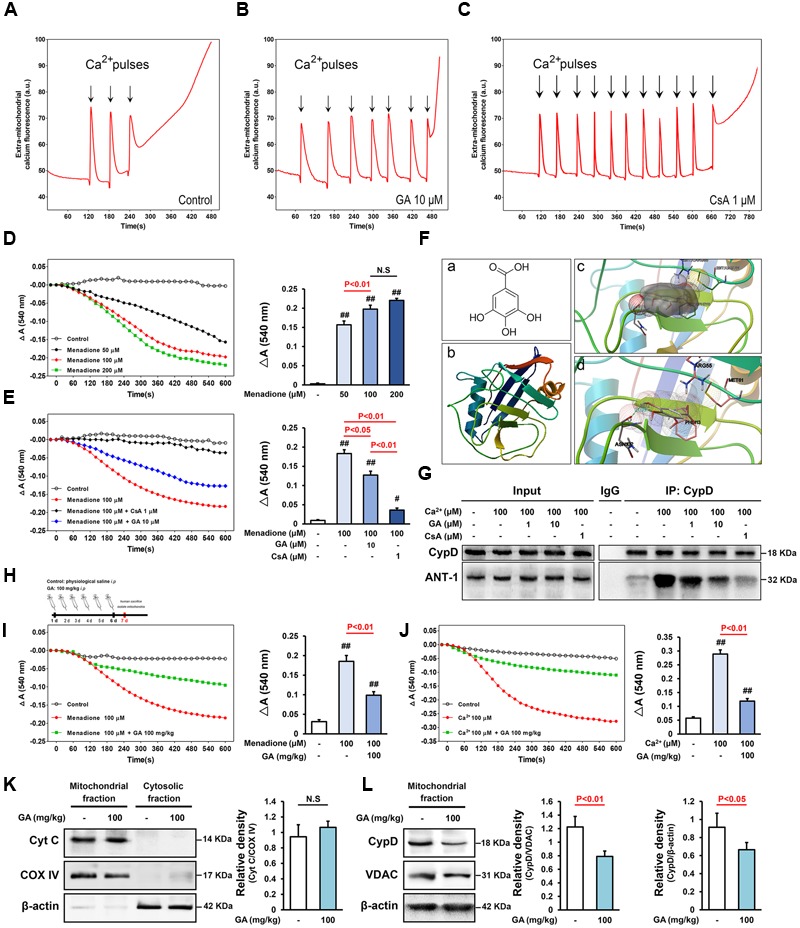
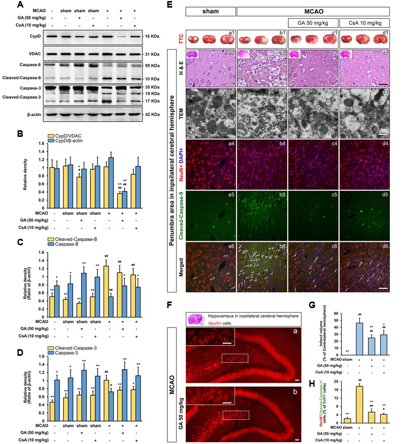
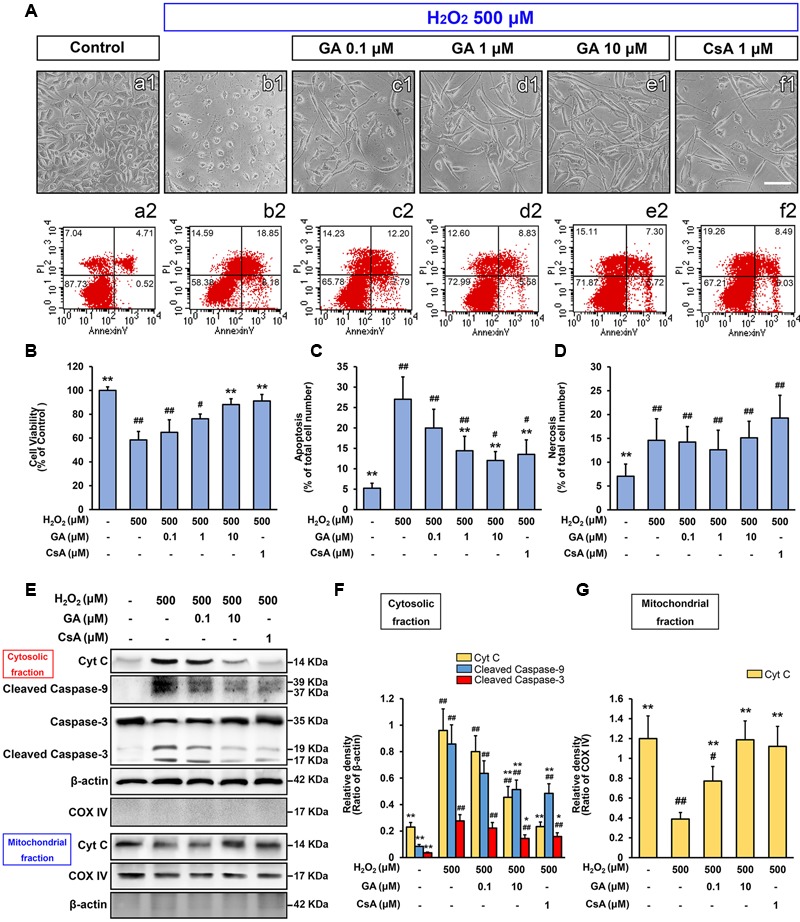
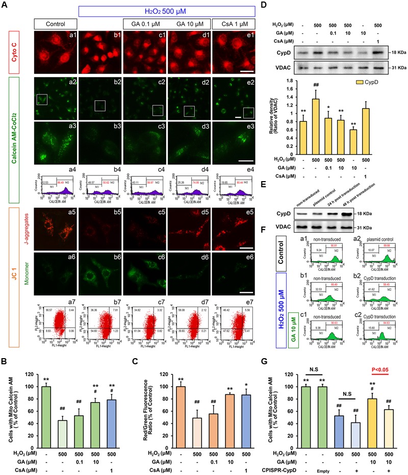
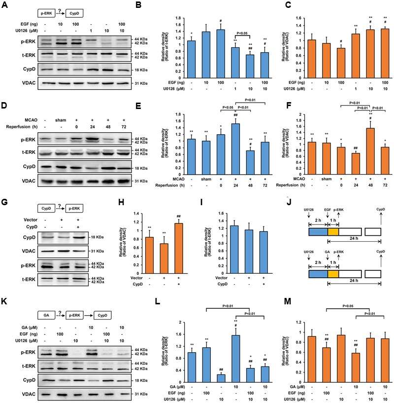
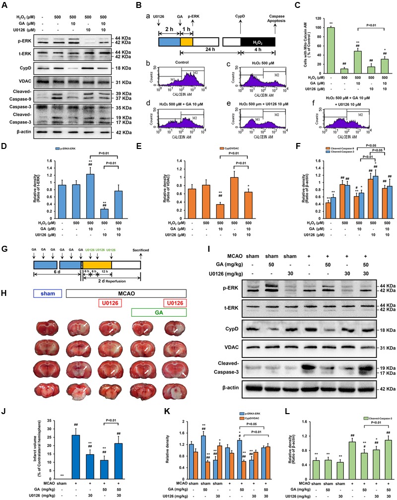
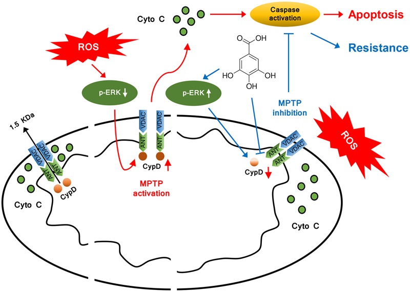
Ischemic stroke is a devastating disease with complex pathophysiology. Much evidence confirms that opening of the mitochondrial permeability transition pore (MPTP) is related with mitochondrial dysfunction to apoptosis in ischemic stroke, thus elucidating its signaling mechanism and screening novel MPTP inhibitor is therefore of paramount importance. Our earlier studies identified that gallic acid (GA), a naturally occurring plant phenol, endows with effect on inhibition of mitochondrial dysfunction, which has significant neuroprotective effect in cerebral ischemia/reperfusion injury. However, its molecular mechanisms regulating mitochondrial dysfunction remain elusive. Here, we uncover a role of GA in protecting mitochondria via MPTP inhibition. In addition to inhibit CypD binding to adenine nucleotide translocator, GA potentiates extracellular signal-regulated kinases (ERK) phosphorylation, leading to a decrease in cyclophilin D (CypD) expression, resulting in a desensitization to induction of MPTP, thus inhibiting caspase activation and ultimately giving rise to cellular survival. Our study firstly identifies ERK-CypD axis is one of the cornerstones of the cell death pathways following ischemic stroke, and confirms GA is a novel inhibitor of MPTP, which inhibits apoptosis depending on regulating the ERK-CypD axis.

Keywords: cerebral ischemia/reperfusion; cyclophilin D (CypD); extracellular signal-regulated kinases (ERK); gallic acid (GA); mitochondrial permeability transition pore (MPTP).


Figures

FIGURE 1
FIGURE 1
Gallic acid (GA) desensitizes MPTP via a signaling axis that involves CypD in liver mitochondria. CRC was determined by the concentration of calcium required to trigger MPTP opening.(A,B) GA has a higher Ca2+ threshold than control mitochondria. (C) CsA (1 μM) robustly increases the number of spikes, consistent with the importance of MPTP in this process. (D) Menadione caused marked mitochondria swelling in a concentration-dependent manner, (E) whereas GA or CsA pre-incubated mitochondria were significantly less sensitive. Histogram comparing ΔA540 values (A540 maxA540 min) among groups (n = 10). (F-a) Chemical structure of GA. (F-b) The 3D structure of CypD obtained from Protein Data Bank (PDB ID: 2BIT). (F-c) Salt bridges to ASN 102, PHE 113, MET 61, ARG 55, and (F-d) an H-bond to ASN 102 make major contributions to the binding affinity for GA (Distance: 2.039, Estimated free energy of binding: –0.568). (G) Representative Immunoprecipitation analysis showed that Ca2+-induced increase of CypD binding to ANT-1 was blocked by CsA or GA (n = 6). GA desensitized liver mitochondria to the permeability transition via suppressing CypD expression. (H) Mitochondria isolated from mouse liver which pre-treatment with GA (100 mg/kg) once a day for 6 days. The level of mitochondrial swelling triggered by menadione (I) or Ca2+ (J) was significantly decreased following GA pre-treatment (n = 10). The release of Cyto C (K), and the expression of CypD (L) were tested via Western Blotting (n = 6). COX IV, VDAC, and β-actin were used as loading controls. Data reported as the means ± SD. P values were obtained using two-way analysis of variance (ANOVA) test. N.S. indicates a P value > 0.05. ##P < 0.01, #P < 0.05 versus control group.
FIGURE 2
FIGURE 2
Gallic acid protects neuron against MCAO insult by the mitochondrial-dependent pathway. GA downregulated brain mitochondrial CypD, inhibited the expression of Cleaved-caspase-8, and Cleaved-caspase-3 protein levels in brain mitochondria following transient cerebral ischemia (n = 4; A–D). VDAC, and β-actin were used as loading controls. GA protected brain mitochondrial and confers neuron survival after transient cerebral ischemia. (E-a1–d1) Representative TTC-stained coronal brain sections; white indicates infarcted tissue. (E-a2–d2) Representative H & E stained coronal brain sections (n = 4). Scale bars, 10 μm. (E-a3–d3) The TEM results evidence that GA protected brain mitochondria against MCAO injury (n = 4). Scale bars, 1 μm. (E-a46, b46, c46, d46) Immunohistochemistry depicting colocalization of Cleaved-caspase-9 with NeuN following 48 h reperfusion. Scale bars, 10 μm. (F) The numbers of pyramidal neurons in the hippocampus region were also diminished by Immunohistochemistry. Scale bars, 10 μm. (G) Histogram showing the infarct volume (% of contralateral hemisphere) in TTC-stained brain sections (n = 8). (H) Histogram showing the percentage of NeuN and Cleaved-caspase-9-positive cells which described as the number of positive cells/total number of cells (n = 4). Data reported as the means ± SD. P values were obtained using two-way analysis of variance (ANOVA) test. ##P < 0.01, #P < 0.05 versus sham group. ∗∗P < 0.01, P < 0.05 versus MCAO group.
FIGURE 3
FIGURE 3
Gallic acid inhibits H2O2-induced SH-SY5Ys death by modulating the intrinsic pathway. The mortality and apoptosis of SH-SY5Y cells were considerably less following pretreated with GA. (A-a2–f2) The number of apoptosis cells were detected by flow cytometry, in accordance with the morphological observations (A-a1–f1). Histogram showing the percentage of cell viability (B), apoptosis (C) and necrosis cells (D), which described as the number of positive cells/total number of cells (n = 5). GA inhibited the expression of mitochondrial apoptotic signaling molecules induced by H2O2. Western Blotting (E), and quantification (F,G) the relative intensities of the bands in each sample by quantity software (n = 4). Scale bars, 5 μm. Data reported as the means ± SD. P values were obtained using two-way analysis of variance (ANOVA) test. ##P < 0.01, #P < 0.05 versus control group. ∗∗P < 0.01, P < 0.05 versus H2O2 group.
FIGURE 4
FIGURE 4
Gallic acid desensitizes the MPTP induced by H2O2 in a CypD-dependent manner. GA decreased the sensitivity of H2O2-induced MPTP induction in SH-SY5Y cells. (A-a1–e1) Representative endogenous Cyto C release in SH-SY5Y cells by Immunofluorescence analysis. (A-a2–e2) MPTP determined by Calcein-CoCl2 fluorescence. (A-a3–e3) Higher magnification and quantification (A-a4–e4) the calcein fluorescence tested by flow cytometry. The representative images show JC-1 aggregates (A-a5–e5), monomers images (A-a6–e6), and quantification (A-a7–e7) the JC-1 by flow cytometry, respectively. Histogram showing the level of calcein fluorescence (B), and the ratio of JC-1 aggregates to JC-1 monomers (C; n = 4). Therapeutic effect of GA on MPTP inhibition was reversed by overexpression of CypD in SH-SY5Y cells. (D) Western blotting (upper) and quantification (lower) for mitochondrial CypD in SH-SY5Y cells pre-treated with GA or CsA following H2O2 for 4 h (n = 4). (E) Transfection efficiency was examined with Western Bolting. VDAC was used to loading control. (F-a1,a2) The levels of calcein fluorescence transfected with CypD or plasmid control, and treated with (F-c1,c2) or without (F-b1,b2) GA under H2O2 conditions were (G) quantificated by flow cytometry (n = 4). Scale bars, 5 μm. Data reported as the means ± SD. P values were obtained using two-way analysis of variance (ANOVA) test. N.S. indicates a P value > 0.05. ##P < 0.01, #P < 0.05 versus control group. ∗∗P < 0.01, P < 0.05 versus H2O2 group.
FIGURE 5
FIGURE 5
Gallic acid alters CypD expression results in an enhancement of ERK phosphorylation. The expression of CypD in SH-SY5Y cells partly depends on ERK phosphorylation. (A) the phosphorylation of ERK and the expression of CypD were determined by immunoblotting at 1 and 24 h following GA or EGF treatment, respectively. t-ERK and VDAC were used as a loading control. (B,C) Histogram showing the relative intensities of the bands in each sample was semi-quantification by quantity software (n = 4). ##P < 0.01, #P < 0.05 versus control group. ∗∗P < 0.01, P < 0.05 versus EGF (100 ng) group. (D–F) Transient cerebral ischemia increases ERK activity and downregulates CypD. The methods of tested and calculated the level of p-ERK and CypD were identical to (A; n = 4). ##P < 0.01, #P < 0.05 versus sham group. ∗∗P < 0.01, P < 0.05 versus Reperfusion 24 h group. CypD was a downstream target protein of ERK. (G) Transfection efficiency was examined with Western bolt. (H,I) The phosphorylation of ERK and the expression of CypD were determined by immunoblotting (n = 4). (J) The design for the in vitro experiment in (K–M) GA altered CypD expression through potentiating ERK phosphorylation. The methods of tested and calculated the level of p-ERK and CypD were identical to (A; n = 4).##P < 0.01, #P < 0.05 versus control group. ∗∗P < 0.01, P < 0.05 versus U0126 (10 μM) group. Data reported as the means ± SD. P values were obtained using two-way analysis of variance (ANOVA) test.
FIGURE 6
FIGURE 6
Extracellular signal-regulated kinases mediates GA-induced cytoprotective effect via CypD downregulation. ERK is necessary for GA to prevent H2O2-induced apoptosis via CypD downregulation. (A) The phosphorylation of ERK and the expression of CypD were determined by immunoblotting. t-ERK, VDAC, and β-actin were used as a loading control. The design for the in vitro experiment in this figure was shown in (B-a). (B-b–f) MPTP determined by Calcein-CoCl2 assay and (C) quantification by flow cytometry (n = 4). (D–F) Histogram showing the relative intensities of the bands in each sample was semi-quantification by quantity software (n = 4). ##P < 0.01, #P < 0.05 versus control group. ∗∗P < 0.01, P < 0.05 versus H2O2 group. ERK signaling triggered by GA were involved in MCAO-mediated apoptosis in rats. (G) The design for the in vivo experiment in this figure. (H) Representative TTC-stained coronal brain sections; white indicates infarcted tissue. (I) The phosphorylation of ERK and the expression of CypD were determined by immunoblotting. (J) Histogram showing the infarct volume (% of contralateral hemisphere) in TTC-stained brain sections (n = 8). (K,L) Quantification the bands in each sample was semi-quantification by quantity software (n = 4). ##P < 0.01, #P < 0.05 versus sham group. ∗∗P < 0.01, P < 0.05 versus MCAO group. Data reported as the means ± SD. P values were obtained using two-way analysis of variance (ANOVA) test.
FIGURE 7
FIGURE 7
Model illustrating the mechanism of cytoprotective effect by GA. In addition to inhibit CypD binding to ANT, GA potentiates ERK phosphorylation, leading to a decrease in CypD expression, resulting in a desensitization to induction of MPTP, thus inhibiting caspase activation and ultimately giving rise to cellular survival.

Similar articles

Cited by

References

    1. Andreux P. A., Houtkooper R. H., Auwerx J. (2013). Pharmacological approaches to restore mitochondrial function. Nat. Rev. Drug Discov. 12 465–483. 10.1038/nrd4023 - DOI - PMC - PubMed
    1. Baines C. P., Kaiser R. A., Purcell N. H., Blair N. S., Osinska H., Hambleton M. A., et al. (2005). Loss of cyclophilin D reveals a critical role for mitochondrial permeability transition in cell death. Nature 434 658–662. 10.1038/nature03434 - DOI - PubMed
    1. Baines C. P., Kaiser R. A., Sheiko T., Craigen W. J., Molkentin J. D. (2007). Voltage-dependent anion channels are dispensable for mitochondrial-dependent cell death. Nat. Cell Biol. 9 550–555. 10.1038/ncb1575 - DOI - PMC - PubMed
    1. Bartsch J. A., Teare G. F., Neufeld A., Hudema N., Muhajarine N. (2013). Secondary prevention of stroke in Saskatchewan, Canada: hypertension control. Int. J. Stroke 8(Suppl. A100) 32–38. 10.1111/j.1747-4949.2012.00930.x - DOI - PubMed
    1. Baumgartner H. K., Gerasimenko J. V., Thorne C., Ferdek P., Pozzan T., Tepikin A. V., et al. (2009). Calcium elevation in mitochondria is the main Ca2+ requirement for mitochondrial permeability transition pore (mPTP) opening. J. Biol. Chem. 284 20796–20803. 10.1074/jbc.M109.025353 - DOI - PMC - PubMed

LinkOut - more resources