Review
. 2017 Apr 6:8:494.
doi: 10.3389/fpls.2017.00494. eCollection 2017.

Non-canonical Translation in Plant RNA Viruses

Affiliations
Review

Non-canonical Translation in Plant RNA Viruses

Manuel Miras et al. Front Plant Sci. .

Abstract

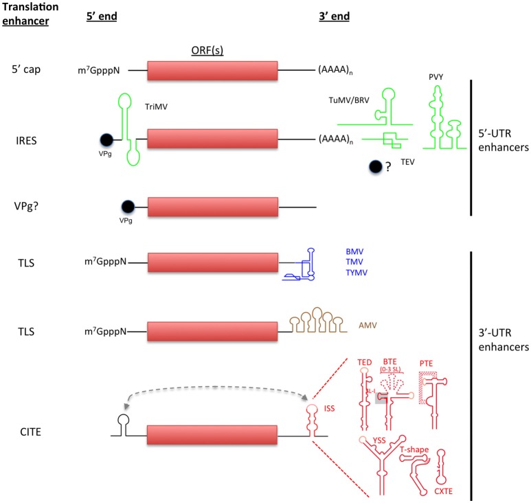
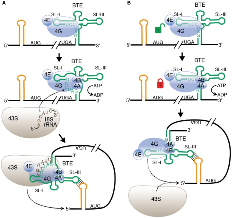
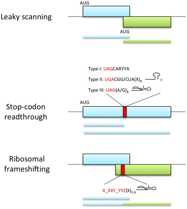
Viral protein synthesis is completely dependent upon the host cell's translational machinery. Canonical translation of host mRNAs depends on structural elements such as the 5' cap structure and/or the 3' poly(A) tail of the mRNAs. Although many viral mRNAs are devoid of one or both of these structures, they can still translate efficiently using non-canonical mechanisms. Here, we review the tools utilized by positive-sense single-stranded (+ss) RNA plant viruses to initiate non-canonical translation, focusing on cis-acting sequences present in viral mRNAs. We highlight how these elements may interact with host translation factors and speculate on their contribution for achieving translational control. We also describe other translation strategies used by plant viruses to optimize the usage of the coding capacity of their very compact genomes, including leaky scanning initiation, ribosomal frameshifting and stop-codon readthrough. Finally, future research perspectives on the unusual translational strategies of +ssRNA viruses are discussed, including parallelisms between viral and host mRNAs mechanisms of translation, particularly for host mRNAs which are translated under stress conditions.

Keywords: 3′-CITE; IRES; RNA structure and function; non-canonical translation; protein synthesis; translation enhancers; translational recoding.


Figures

Figure 1
Figure 1
Non-canonical initiation translation mechanisms used by plant RNA viruses. Canonical translation of eukaryotic mRNAs is shown in the top. Non-canonical translation elements are grouped depending on their location in viral genome and are color-coded to match with the virus acronyms. Lighter-shaded loops in the secondary structure of 3′-CITEs indicated sequences known or predicted to base-pair to the 5′ end of the viral genome (shown as dashed line).
Figure 2
Figure 2
Alternative models of ribosome recruitment and delivery to the 5′-UTR via the BTE. (A) Base pairing to rRNA model. Top: eIF4F binds to SL-I of the BTE (green) through the eIF4G subunit. eIF4E enhances but is not required for BTE binding. Middle: Helicase (eIF4A + eIF4B) binds and uses ATP hydrolysis to unwind GAUCCU, making it available to base pair to 18S rRNA at a conserved sequence in the region where the Shine-Dalgarno binding site is located in prokaryotic 16S rRNA. Bottom: The 43S preinitiation complex base pairs to the BTE and is delivered to the 5′ end by long-distance base pairing (yellow stem-loop). (B) Conventional ribosome recruitment model. Top: eIF4F binds BTE as in (A). Middle: Binding of eIF4A + eIF4B and ATP hydrolysis increases binding affinity of eIF4F, “locking” it on to the BTE, perhaps by altering the structure of BTE RNA. Bottom: eIF4 complex is delivered to 5′ end by long-distance base pairing where it recruits the 43S preinitiation complex to the RNA. In both models, 43S scanning from the 5′ end to the start codon is the same as in normal cap-dependent translation. Not shown: other factors, such as eIF3 and factors in the preinitiation complex.
Figure 3
Figure 3
Viral recoding strategies. Top panel represents leaky scanning mechanism where ribosomes fail to start translation at the first AUG codon and continue scanning until they reach an alternative start codon in the optimal initiation context. This process allows the expression of two proteins with distinct amino acid sequence when the initiation sites are in different reading frames (as shown) or C-terminally coincident isoforms of a single protein if initiation sites are in-frame (not shown). Middle panel shows the expression of proteins with alternative C-terminal because a portion of ribosomes fail to terminate at a stop codon and continue translation. The efficiency of readthrough can be stimulated by the presence of elements downstream of the stop codon: UAG stop codon followed by the consensus motif CARYYA, where R is a purine and Y is a pyrimidine (Type I); UGA stop codon followed by CGG or CUA triplet and a stem-loop structure separated from the stop codon by 8 nt (Type II); UAG stop codon and adjacent G or purine octanucleotide and a compact pseudoknot structure (Type III). Bottom panel represents ribosomal frameshifting strategy, where ribosomes are directed into a different reading frame guided by the slippery signal X_XXY_YYZ (X and Y can be any base and Z is any base except G) and a secondary structure element located 5-9 nt downstream the slippery sequence.

References

    1. Adams M. J., Antoniw J. F., Beaudoin F. (2005). Overview and analysis of the polyprotein cleavage sites in the family Potyviridae. Mol. Plant Pathol. 6, 471–487. 10.1111/j.1364-3703.2005.00296.x - DOI - PubMed
    1. Afonina Z. A., Myasnikov A. G., Shirokov V. A., Klaholz B. P., Spirin A. S. (2014). Formation of circular polyribosomes on eukaryotic mRNA without cap-structure and poly (A)-tail: a cryo electron tomography study. Nucleic Acids Res. 42, 9461–9469. 10.1093/nar/gku599 - DOI - PMC - PubMed
    1. Agranovsky A. A., Koonin E. V., Boyko V. P., Maiss E., Frötschl R., Lunina N. A., et al. . (1994). Beet yellows closterovirus: complete genome structure and identification of a leader papain-like thiol protease. Virology 198, 311–324. 10.1006/viro.1994.1034 - DOI - PubMed
    1. Aitken C. E., Lorsch J. R. (2012). A mechanistic overview of translation initiation in eukaryotes. Nat. Struct. Mol. Biol. 19, 568–576. 10.1038/nsmb.2303 - DOI - PubMed
    1. Annamalai P., Rao A. L. (2007). In vivo packaging of brome mosaic virus RNA3, but not RNAs 1 and 2, is dependent on a cis-acting 3′ tRNA-like structure. J. Virol. 81, 173–181. 10.1128/JVI.01500-06 - DOI - PMC - PubMed