. 2017 Apr 17:13:23.
doi: 10.1186/s13223-017-0194-9. eCollection 2017.

Regulation of Th2 responses by different cell types expressing the interleukin-31 receptor

Affiliations

Regulation of Th2 responses by different cell types expressing the interleukin-31 receptor

Saburo Saito et al. Allergy Asthma Clin Immunol. .

Abstract

Background: Interleukin-31 (IL-31) is a recently identified cytokine produced by Th2 cells that is involved in the development of atopic dermatitis-induced skin inflammation and pruritus. Its receptor, IL-31RA, is expressed by a number of cell types, including epithelial cells, eosinophils, and activated monocytes and macrophages. To date, however, the regulation of Th2 responses by distinct cell types and tissues expressing IL-31RA has not been well studied.

Methods: In this study, Cry j 2, one of the major allergens of Japanese cedar pollen, was administered to IL-31RA-deficient or wild-type (WT) mice via nasal or intraperitoneal injection for induction of specific Th2 responses.

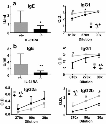
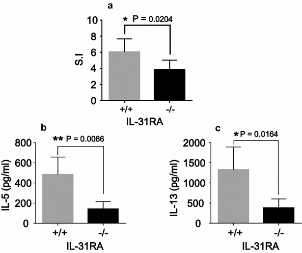
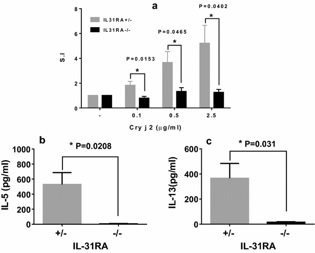
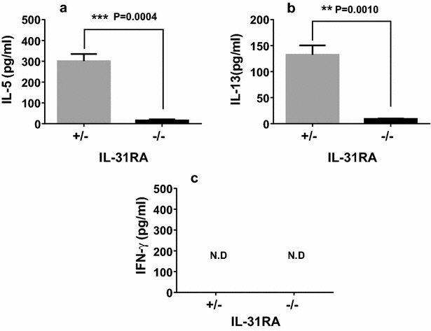
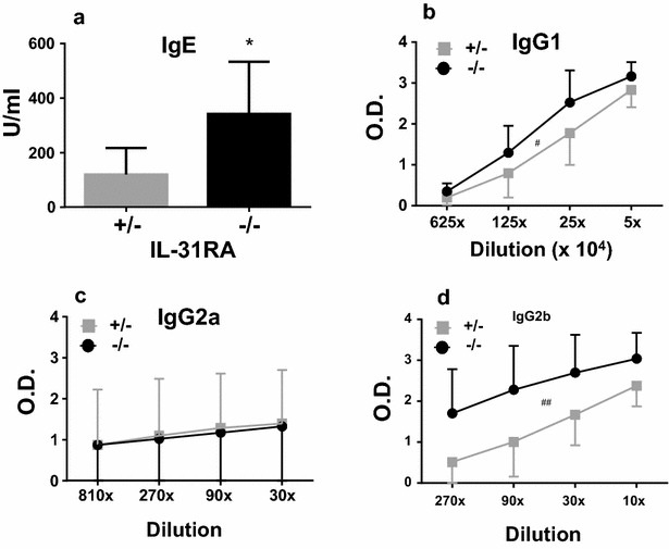
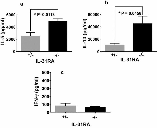
Results: After nasal administration of Cry j 2, IL-31RA-deficient mice showed lower Cry j 2-specific CD4+ T cell proliferation, Th2 cytokine (IL-5 and IL-13) production, and Th2-mediated (IgE, IgG1, and IgG2b) antibody responses than WT mice. In contrast, IL-31RA-deficient mice administered Cry j 2 intraperitoneally showed stronger Th2 immune responses than WT mice.

Conclusions: These results indicate that IL-31R signaling positively regulates Th2 responses induced by nasal administration of Cry j 2, but negatively regulates these responses when Cry j 2 is administered intraperitoneally. Collectively, these data indicate that the induction of antigen-specific Th2 immune responses might depend on tissue-specific cell types expressing IL-31RA.

Keywords: Cry j 2; Deficient mice; IL-31 receptor; IgE; Th2.


Figures

Fig. 1
Fig. 1
Generation of IL-31RA-deficient mice. To generate C57BL/6-IL-31RAtLacZ/+ knock-out (KO) mice, we used homologous recombination in embryonic stem (ES) cells to create a mutant allele in which exon 4 of the IL-31RA gene was replaced by a cassette expressing the selective marker neomycin transferase (a). Two recombinant ES cells were found to be IL-31RAtm1(LacZ). Chimeric mice were mated with C57BL/6 mice to produce mutant IL-31RA+/LacZ(+/−) progeny. The generation of mutant IL-31RA+/− mice was verified by Southern blot analysis (b). The IL-31RA+/− allele was then backcrossed to C57BL/6 mice for 15 more generations using male IL-31RA+/−. Homozygous IL-31RA−/− and WT (IL-31RA+/+) littermates were generated by intercrossing IL-31RA+/− mice from the 15th generations of backcrossed mice. Heterozygous IL-31RA+/− and homozygous IL-31RA−/− littermates were generated by crossing IL-31RA+/− mice with homozygous IL-31RA−/− mice. The expression of IL-31RA or beta-galactosidase of skin from IL-31RA+/+ and IL-31RA−/− mice was revealed by immunohistological staining with antibodies against each antigen (c)
Fig. 2
Fig. 2
IL-31RA−/− mice nasally injected with Cry j 2 exhibit decreased antigen-specific Th2-mediated antibody responses. Serum from WT (IL-31RA+/+, N = 6) and KO (IL-31RA−/−, N = 7) mice following 15 nasal administrations of Cry j 2 was assayed for antigen-specific IgE and IgG1 antibodies (a). Littermate IL-31RA−/− mice (N = 15) and IL-31RA+/− control mice (N = 10) were nasally administered Cry j 2 antigen five times a week for induction of specific Th2 responses. Serum from littermate mice following 15 nasal administrations was assayed for antigen-specific IgE, IgG1, IgG2a, and IgG2b levels (b) by ELISA. The data shown are of one experiment representative of three independent experiments. Data are presented as the mean ± SD. Unpaired t test; *P < 0.05. **P < 0.01
Fig. 3
Fig. 3
IL-31RA−/− mice nasally injected with Cry j 2 show decreased antigen-specific proliferative and Th2 cytokine responses. 7 days after 15 nasal administrations of Cry j 2, spleen cells from WT (IL-31RA+/+, N = 6) and KO (IL-31RA−/−, N = 7) mice were cultured for 88 h in the presence or absence of Cry j 2 (0.1 μg/ml) and pulsed for the last 16 h with 0.5 μCi of tritiated thymidine. The results of T-cell proliferation (a) are expressed in terms of a stimulation index (SI) calculated by dividing counts per minute (cpm) in the presence of antigen with cpm in the absence of antigen. Supernatants obtained from a 72-h cell culture were assayed by ELISA for IL-13 (b) and IL-5 (c). Data are presented as the mean ± SD
Fig. 4
Fig. 4
Decreased antigen-specific proliferative and Th2 cytokine responses of spleen cells from IL-31RA−/− mice nasally injected with Cry j 2. Seven days after 19 nasal administrations of Cry j 2, spleen cells from six representative IL-31RA−/− and littermate control IL-31RA+/− mice were cultured for 88 h in the presence or absence of the indicated dose of Cry j 2 and pulsed for the last 16 h with 0.5 μCi of tritiated thymidine to analyze antigen-specific T-cell proliferative responses (a). Supernatants from 72-h cell cultures were assayed for IL-13 (b) and IL-5 (c). Data are presented as the mean ± SD
Fig. 5
Fig. 5
Decreased antigen-specific Th2 cytokine responses of submandibular lymph node cells from IL-31RA−/− mice nasally injected with Cry j 2. 7 days after 19 nasal administrations of Cry j 2, submandibular lymph node cells from six representative IL-31RA−/− and littermate control IL-31RA+/− mice were pooled and cultured. Supernatants from a 72-h cell culture were assayed by ELISA for IL-5 (a), IL-13 (b), and IFN-γ (c). Data are presented as the mean ± SD
Fig. 6
Fig. 6
IL-31RA−/− mice administered intraperitoneally with Cry j 2 exhibit enhanced antigen-specific Th2-mediated antibody responses. Littermate IL-31RA−/− and IL-31RA+/− control mice were intraperitoneally administered once a week with Cry j 2 for induction of antigen-specific Th2 responses. 7 days after three intraperitoneal administrations of Cry j 2, serum from IL-31RA−/− (N = 5) and littermate control IL-31RA+/− (N = 5) mice were collected and assayed for Cry j 2-specific IgE (a), IgG1 (b), IgG2a (c), and IgG2b (d) antibody responses. Data are presented as the mean ± SD. Unpaired t test; *P < 0.05. Paired t test; #P < 0.05, ##P < 0.01
Fig. 7
Fig. 7
IL-31RA−/− mice intraperitoneally administered Cry j 2 show enhanced antigen-specific proliferative and Th2 cytokine responses. For the analysis of Th2 cytokine responses, spleen cells were collected from mice 7 days after five intraperitoneal administrations of Cry j 2 and cultured with Cry j 2. Supernatants from 72-h cell cultures were assayed for IL-5 (a), IL-13 (b), and IFN-γ (c). The data shown are of one experiment representative of five independent experiments with 4 to 10 mice per group. Data are presented as the mean ± SD

Similar articles

Cited by

References

    1. Dillon SR, Sprecher C, Hammond A, Bilsborough J, Rosenfeld-Franklin M, Presnell SR, et al. Interleukin 31, a cytokine produced by activated T cells, induces dermatitis in mice. Nat Immunol. 2004;5(7):752–760. doi: 10.1038/ni1084. - DOI - PubMed
    1. Bilsborough J, Leung DY, Maurer M, Howell M, Boguniewicz M, Yao L, et al. IL-31 is associated with cutaneous lymphocyte antigen-positive skin homing T cells in patients with atopic dermatitis. J Allergy Clin Immunol. 2006;117(2):418–425. doi: 10.1016/j.jaci.2005.10.046. - DOI - PubMed
    1. Ezzat MH, Hasan ZE, Shaheen KY. Serum measurement of interleukin-31 (IL-31) in paediatric atopic dermatitis: elevated levels correlate with severity scoring. J Eur Acad Dermatol Venereol. 2011;25(3):334–339. doi: 10.1111/j.1468-3083.2010.03794.x. - DOI - PubMed
    1. Neis MM, Peters B, Dreuw A, Wenzel J, Bieber T, Mauch C, et al. Enhanced expression levels of IL-31 correlate with IL-4 and IL-13 in atopic and allergic contact dermatitis. J Allergy Clin Immunol. 2006;118(4):930–937. doi: 10.1016/j.jaci.2006.07.015. - DOI - PubMed
    1. Perrigoue JG, Li J, Zaph C, Goldschmidt M, Scott P, de Sauvage FJ, et al. IL-31–IL-31R interactions negatively regulate type 2 inflammation in the lung. J Exp Med. 2007;204(3):481–487. doi: 10.1084/jem.20061791. - DOI - PMC - PubMed