. 2017 Apr 19:13:29.
doi: 10.1186/s13007-017-0180-8. eCollection 2017.

Leucine-rich-repeat-containing variable lymphocyte receptors as modules to target plant-expressed proteins

Affiliations

Leucine-rich-repeat-containing variable lymphocyte receptors as modules to target plant-expressed proteins

André C Velásquez et al. Plant Methods. .

Abstract

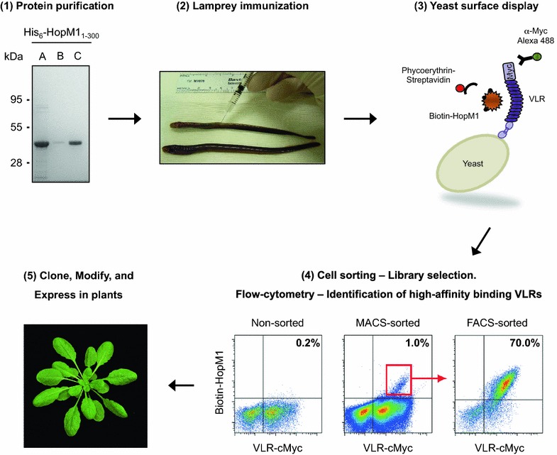
Background: The ability to target and manipulate protein-based cellular processes would accelerate plant research; yet, the technology to specifically and selectively target plant-expressed proteins is still in its infancy. Leucine-rich repeats (LRRs) are ubiquitously present protein domains involved in mediating protein-protein interactions. LRRs confer the binding specificity to the highly diverse variable lymphocyte receptor (VLR) antibodies (including VLRA, VLRB and VLRC types) that jawless vertebrates make as the functional equivalents of jawed vertebrate immunoglobulin-based antibodies.

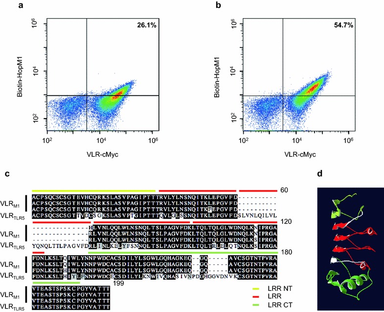
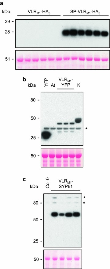
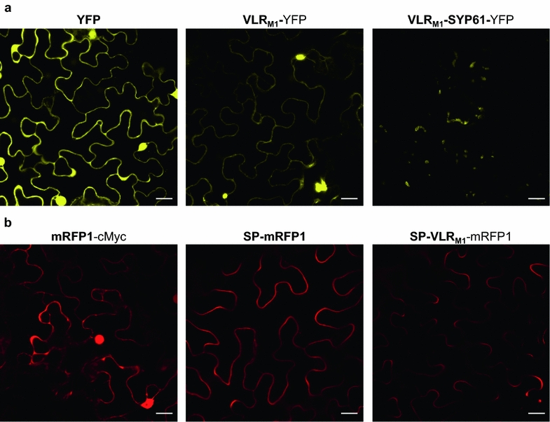
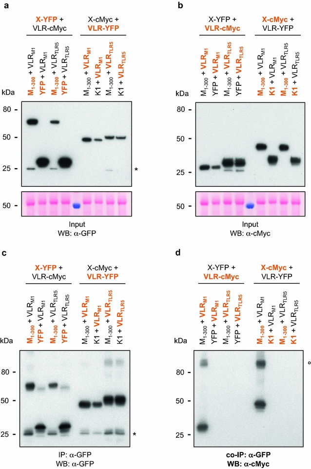
Results: In this study, VLRBs targeting an effector protein from a plant pathogen, HopM1, were developed by immunizing lampreys and using yeast surface display to select for high-affinity VLRBs. HopM1-specific VLRBs (VLRM1) were expressed in planta in the cytosol, the trans-Golgi network, and the apoplast. Expression of VLRM1 was higher when the protein localized to an oxidizing environment that would favor disulfide bridge formation (when VLRM1 was not localized to the cytoplasm), as disulfide bonds are necessary for proper VLR folding. VLRM1 specifically interacted in planta with HopM1 but not with an unrelated bacterial effector protein while HopM1 failed to interact with a non-specific VLRB.

Conclusions: In the future, VLRs may be used as flexible modules to bind proteins or carbohydrates of interest in planta, with broad possibilities for their use by binding directly to their targets and inhibiting their action, or by creating chimeric proteins with new specificities in which endogenous LRR domains are replaced by those present in VLRs.

Keywords: HopM1; Leucine-rich repeat; Modules; Protein targeting; Variable lymphocyte receptor.


Figures

Fig. 1
Fig. 1
Variable lymphocyte receptors as tools to target plant-expressed proteins. A schematic diagram depicting the steps involved in developing an LRR-containing VLR that binds to plant-expressed proteins. (1) Express and purify the protein from E. coli, P. pastoris, or other sources. (2) Immunize lampreys with the purified protein of interest conjugated to an adjuvant for the production of VLRB antibodies. (3) Clone VLRBs from lamprey’s lymphocytes into a yeast surface display (YSD) library. (4) Enrich the YSD library for high-affinity binding VLRBs using magnetic-activated cell sorting (MACS) and fluorescence-activated cell sorting (FACS), and identify individual high-affinity binding VLRBs using flow cytometry. (5) Clone VLRBs into plant expression vectors for in planta expression. The LRR-containing VLR may be modified to carry additional modules (e.g., enzymes or receptors). Step 1 shows Denville Blue™ staining of SDS-PAGE gel of E. coli expressed His6-HopM11–300. (A) Ni–NTA agarose purified protein. (B) Anion-exchange chromatographic flow-through. (C) Fraction eluted with 433 mM NaCl from the anion exchange chromatographic column, which after dialysis into phosphate buffer was used to inoculate lampreys for VLRB production. Step 4 shows the YSD library before enrichment for VLRBs that bind HopM11–300 with high affinity (non-sorted), and after MACS and FACS selection
Fig. 2
Fig. 2
Identification of variable lymphocyte receptors that bind the Pseudomonas syringae effector HopM1. a and b yeast-surface display of HopM1-specific VLRBs. The x-axis represents Alexa Fluor® 488 (conjugated to α-c-Myc antibody) fluorescence while the y-axis shows phycoerythrin (conjugated to streptavidin) fluorescence of individual yeast cells. Fluorescence was detected using BD Accuri C6 flow cytometer. The number highlighted in bold indicates the percentage of yeast cells with detectable HopM11–300 binding. a Lower-affinity binding HopM1-specific VLRB; 50 nM biotinylated HopM1. b Higher-affinity binding HopM1-specific VLRB; 50 nM biotinylated HopM1. c Amino acid alignment of the three different high-affinity HopM1-specific VLRB sequences and of Toll-like receptor 5 (TLR5; a mammalian immune receptor that recognizes bacterial flagellin)-specific VLRB. Alignment was generated using MegAlign (DNASTAR) and graphed using Boxshade. Highlighted in a white background are amino acids that are different. Amino acid position is shown on the upper right corner. A red bar over the sequence identifies the leucine-rich repeat (LRR) domains identified. In yellow and green, are the N-terminal and C-terminal LRR domains, respectively, which are characterized by the presence of disulfide bonds. The domains were identified using the SMART tool [59]. d 3-D structure model of HopM1-specific VLRB. LRR domains are highlighted in red, the N-terminal LRR domain in yellow, and the C-terminal LRR domain in green. Modeling was performed with SWISS-MODEL using the structure of a previously crystalized VLRB protein (3g3aA) [16]
Fig. 3
Fig. 3
In planta accumulation of VLRM1 is higher if the protein goes through the secretory pathway. a Western blot of cytoplasmic and apoplastic VLRBs fused at their C-terminus with three HA tags detected with α-HA antibodies. Eight µg of total protein were loaded per well. Expression of three different high-affinity HopM1-specific VLRBs (done in duplicate) with slightly different amino acid sequences (see Fig. 2c) was performed for the cytoplasmic (VLRM1-HA3) and apoplastic (SP-VLRM1-HA3) versions of the HopM1-specific VLRB. A Ponceau S staining of the membrane is shown below the blot to confirm similar sample loading of the gel. Accumulation of only SP-VLRM1 was observed. SP signal peptide. b Western blot of cytoplasmic VLRB fused at its C-terminus with YFP detected with α-GFP antibodies. Twenty-nine µg of total protein were loaded per sample. Expression of three different high-affinity HopM1-specific VLRBs with slightly different amino acid sequences was performed. YFP and HopK1-YFP (K) were used as positive controls, while an A. tumefaciens strain (At) devoid of any plant expression vectors was used as a negative control. The asterisk represents the position of a non-specific band. A Ponceau S staining of the membrane is shown below the blot to confirm similar sample loading of the gel. c Western blot of cytoplasmic VLRB fused at its C-terminus with syntaxin SYP61 (At1g28490) and three HA tags detected with α-HA antibodies. Twenty-three µg of total protein were loaded per well. Proteins were extracted from four T1 transgenic A. thaliana Col-0 plants and an untransformed Col-0 plant. Asterisks represent the position of non-specific bands. A Ponceau S staining of the membrane is shown below the blot to confirm similar sample loading
Fig. 4
Fig. 4
Visualization of in planta VLRB protein expression. a Expression of intracellular YFP, HopM1-specific VLRB (VLRM1), and VLRM1 fused to A. thaliana syntaxin SYP61 (At1g28490) in Nicotiana benthamiana. Images were taken with the Olympus FluoView 1000 confocal microscope using a 515 nm laser for YFP excitation, while emission was collected between 530 and 569 nm. Fusing VLRM1 to SYP61 targets the VLRB to intracellular compartments, most likely the trans-Golgi network. b Expression of intracellular mRFP1, and predicted extracellular SP-mRFP1 and SP-VLRM1-mRFP1 in N. benthamiana. mRFP1 and HopM1-specific VLRB were targeted to the apoplast by fusing the VLRB to the signal peptide (SP) of Arabidopsis thaliana PR1 (At2g14610). Accumulation on the periphery of the cells was observed. Images were taken with the Olympus FluoView 1000 confocal microscope using a 559 nm laser for excitation and collecting the emission between 570 and 600 nm. White bar length represents 20 µm. Image brightness increased 40%
Fig. 5
Fig. 5
In planta interaction of HopM1 with VLRM1. Co-Immunoprecipitation of HopM1 and its corresponding VLRB in Nicotiana benthamiana. Thirteen and one-half mg of proteins were immunoprecipitated (IP) with α-GFP agarose beads without the use of reducing agents in the buffers. Proteins were detected with either α-GFP (IP) or α-c-Myc (co-IP) antibodies. Interactions between HopM1 and HopM1-specific VLRB were tested with both proteins fused to two different epitope tags. Highlighted in orange are those proteins detected in the Western blot, while in black are those proteins also expressed but not detected. VLRM1 = SP-VLRM1, VLRTLR5 = SP-VLRTLR5, M1–300 = SP-HopM11–300, K1 = HopK1. a Total protein input of YFP-tagged proteins for IP, Western blot used α-GFP antibodies for protein detection. Ponceau S staining of the input PVDF membrane is shown below the Western blot. The asterisk represents the position of YFP cleaved from the fusion protein. b Total protein input of c-Myc-tagged proteins for IP. Western blot used α-c-Myc antibodies for protein detection. Ponceau S staining of the input PVDF membrane is shown below the Western blot. c IP of YFP-tagged proteins with α-GFP agarose beads. Western blot used α-GFP antibodies for protein detection. The asterisk indicates the position of YFP cleaved from the fusion protein. d Co-IP of c-Myc-tagged proteins with α-GFP agarose beads. Western blot used α-c-Myc antibodies for protein detection. VLRM1 only interacted with HopM1. The open circle marks the position of a probable dimer formed between VLRM1 and HopM11–300

Similar articles

Cited by

References

    1. Kobe B, Kajava AV. The leucine-rich repeat as a protein recognition motif. Curr Opin Struct Biol. 2001;11:725–732. doi: 10.1016/S0959-440X(01)00266-4. - DOI - PubMed
    1. Kobe B, Deisenhofer J. A structural basis of the interactions between leucine-rich repeats and protein ligands. Nature. 1995;374:183–186. doi: 10.1038/374183a0. - DOI - PubMed
    1. Jeong S, Trotochaud AE, Clark SE. The Arabidopsis CLAVATA2 gene encodes a receptor-like protein required for the stability of the CLAVATA1 receptor-like kinase. Plant Cell. 1999;11:1925–1934. doi: 10.1105/tpc.11.10.1925. - DOI - PMC - PubMed
    1. Stergiopoulos I, van den Burg HA, Okmen B, Beenen HG, van Liere S, Kema GH, et al. Tomato Cf resistance proteins mediate recognition of cognate homologous effectors from fungi pathogenic on dicots and monocots. Proc Natl Acad Sci USA. 2010;107:7610–7615. doi: 10.1073/pnas.1002910107. - DOI - PMC - PubMed
    1. She J, Han Z, Kim TW, Wang J, Cheng W, Chang J, et al. Structural insight into brassinosteroid perception by BRI1. Nature. 2011;474:472–476. doi: 10.1038/nature10178. - DOI - PMC - PubMed

LinkOut - more resources