Case Reports
. 2017:2017:4695491.
doi: 10.1155/2017/4695491. Epub 2017 Mar 27.

Acute Myeloid Leukemia with Basophilic Differentiation Transformed from Myelodysplastic Syndrome

Affiliations
Case Reports

Acute Myeloid Leukemia with Basophilic Differentiation Transformed from Myelodysplastic Syndrome

Yasuhiro Tanaka et al. Case Rep Hematol. 2017.

Abstract

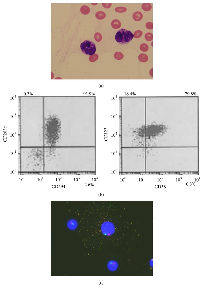
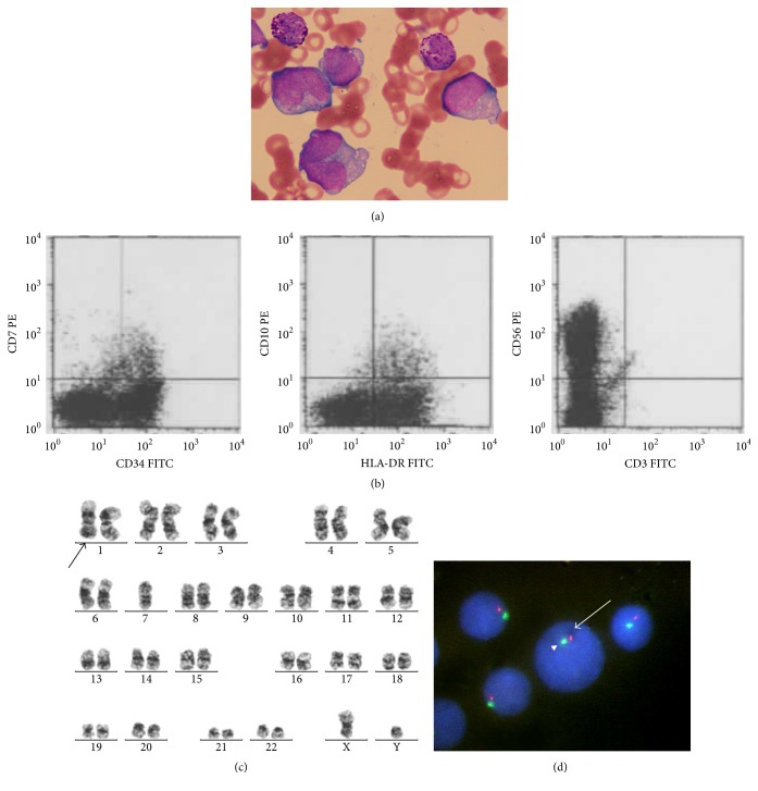
Myelodysplastic syndrome (MDS) terminally transforms to acute myeloid leukemia (AML) or bone marrow failure syndrome, but acute myeloid leukemia with basophilic differentiation has been rarely reported. An 81-year-old man was referred to our department for further examination of intermittent fever and normocytic anemia during immunosuppressive treatment. Chromosomal analysis showed additional abnormalities involving chromosome 7. He was diagnosed as having MDS. At the time of diagnosis, basophils had not proliferated in the bone marrow. However, his anemia and thrombocytopenia rapidly worsened with the appearance of peripheral basophilia three months later. He was diagnosed as having AML with basophilic differentiation transformed from MDS. At that time, monosomy 7 was detected by chromosomal analysis. We found that basophils can be confirmed on the basis of the positivity for CD203c and CD294 by flow cytometric analysis. We also found by cytogenetic analysis that basophils were derived from myeloblasts. He refused any chemotherapy and became transfusion-dependent. He died nine months after the transformation. We should keep in mind that MDS could transform to AML with basophilic differentiation when peripheral basophilia in addition to myeloblasts develops in patients with MDS.


Conflict of interest statement

The authors declare no conflict of interests.

Figures

Figure 1
Figure 1
The characterization of basophils in peripheral blood. (a) Peripheral blood smear showed some medium- to large-sized abnormal cells with lobulated nucleus, and many basophilic granules were observed (May-Giemsa staining, original magnification, ×1000). (b) Flow cytometric analysis showed that abnormal cells were positive for CD38, CD123, CD203c, and CD294, which was consistent with basophils. (c) Dual-color FISH analysis using chromosome 7 probes showed that abnormal cell with many granules contained one red signal and one green signal, which indicated monosomy 7 (D7S486 probe, red signal; D7Z1 probe, green signal). Other cells without any granules also showed monosomy 7.
Figure 2
Figure 2
The characterization of myeloblasts in bone marrow. (a) Bone marrow smear showed some large-sized myeloblasts with clear nucleoli, basophilic cytoplasm, and some vacuoles. Some mature basophils were observed (May-Giemsa staining, original magnification, ×1000). (b) Flow cytometric analysis showed blasts positive for CD34, CD56, and HLA-DR. (c) Chromosomal analysis by G banding showed 45, XY, -7 [5]/45, idem, add(1)(q21) [4]/46, XY [2]. (d) Dual-color FISH analysis using chromosome 7 probes showed that myeloblasts contained one red signal and one green signal, which indicated monosomy 7 (D7S486 probe, red signal, arrow; D7Z1 probe, green signal, arrow head).

Similar articles

Cited by

  • Monoblastic leukemia (M5a) with chronic basophilic leukemia in a cat.
    Shimoda T, Tanabe M, Shoji Y, Kanda T, Kishida K, Kishida A, Hashimoto A, Otsuka M, Miyamoto A, Kawakita T, Oshita W, Hirao R, Suwa A. Shimoda T, et al. J Vet Med Sci. 2022 Feb 23;84(2):251-256. doi: 10.1292/jvms.21-0383. Epub 2021 Dec 15. J Vet Med Sci. 2022. PMID: 34911870 Free PMC article.

References

    1. Brunning R. D., Orazi A., Germing U., et al. WHO Classification of Tumors of Haematopoietic and Lymphoid Tissues. Lyon, France: IARC; 2008. Myelodysplastic syndrome/neoplasms, overview; pp. 88–93.
    1. Matsushima T., Handa H., Yokohama A., et al. Prevalence and clinical characteristics of myelodysplastic syndrome with bone marrow eosinophilia or basophilia. Blood. 2003;101(9):3386–3390. doi: 10.1182/blood-2002-03-0947. - DOI - PubMed
    1. Wimazal F., Germing U., Kundi M., et al. Evaluation of the prognostic significance of eosinophilia and basophilia in a larger cohort of patients with myelodysplastic syndromes. Cancer. 2010;116(10):2372–2381. doi: 10.1002/cncr.25036. - DOI - PubMed
    1. Arber D. A., Brunning R. D., Orazi A., et al. WHO Classification of Tumors of Haematopoietic and Lymphoid Tissues. Lyon, France: IARC; 2008. Acute myeloid leukemia, not otherwise specified; pp. 130–139.
    1. Bahmanyar M., Chang H. Acute myeloid leukemia with myelodysplasia-related changes demonstrating prominent basophilic differentiation. Blood. 2016;127(20, article 2503) doi: 10.1182/blood-2016-02-701144. - DOI - PubMed

Publication types

LinkOut - more resources