Review
. 2017 Apr 6:4:19.
doi: 10.3389/fmolb.2017.00019. eCollection 2017.

Physiological Functions of the Cellular Prion Protein

Affiliations
Review

Physiological Functions of the Cellular Prion Protein

Andrew R Castle et al. Front Mol Biosci. .

Abstract

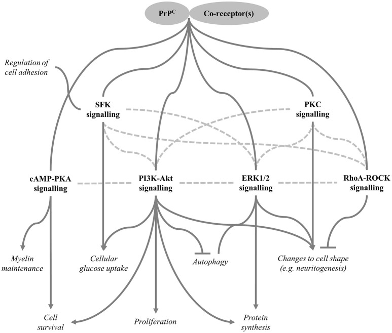
The prion protein, PrPC, is a small, cell-surface glycoprotein notable primarily for its critical role in pathogenesis of the neurodegenerative disorders known as prion diseases. A hallmark of prion diseases is the conversion of PrPC into an abnormally folded isoform, which provides a template for further pathogenic conversion of PrPC, allowing disease to spread from cell to cell and, in some circumstances, to transfer to a new host. In addition to the putative neurotoxicity caused by the misfolded form(s), loss of normal PrPC function could be an integral part of the neurodegenerative processes and, consequently, significant research efforts have been directed toward determining the physiological functions of PrPC. In this review, we first summarise important aspects of the biochemistry of PrPC before moving on to address the current understanding of the various proposed functions of the protein, including details of the underlying molecular mechanisms potentially involved in these functions. Over years of study, PrPC has been associated with a wide array of different cellular processes and many interacting partners have been suggested. However, recent studies have cast doubt on the previously well-established links between PrPC and processes such as stress-protection, copper homeostasis and neuronal excitability. Instead, the functions best-supported by the current literature include regulation of myelin maintenance and of processes linked to cellular differentiation, including proliferation, adhesion, and control of cell morphology. Intriguing connections have also been made between PrPC and the modulation of circadian rhythm, glucose homeostasis, immune function and cellular iron uptake, all of which warrant further investigation.

Keywords: PrPC; adhesion; differentiation; myelin maintenance; prion; proliferation; stress protection; transmissible spongiform encephalopathies.


Figures

Figure 1
Figure 1
Theories of the evolutionary history of the prion gene family. The figure shows three possibilities for the evolution of the mammalian prion gene family. (A) Schmitt-Ulms et al. (2009) proposed that an ancestral PRNP gene evolved from a member of the ZIP metal ion transporter family. Subsequently, this PRNP precursor gave rise to the modern-day prion gene family through local duplications and other genomic rearrangements. (B) This alternative version, also put forward by Schmitt-Ulms et al. (2009), incorporates additional research by Premzl et al. (2004) suggesting that SPRN existed before PRNP and that the genetic material encoding the N-terminal domain of an ancestral PrPC evolved from a gene called SPRNB1 that, itself, had emerged from the original SPRN. The genetic material encoding the C-terminal domain of the ancestral PrPC is proposed to have derived from a ZIP gene and a later, local duplication would have then created modern-day PRNP and PRND. Although, descendants of SPRNB2 are found in fish, this gene is thought either to have been deleted or to have evolved beyond detectability in the mammalian lineage (Premzl et al., 2004). (C) A further possibility is that ancestral PRNP and SRPN genes could have evolved out of ZIP genes in separate events (Westaway et al., 2011).
Figure 2
Figure 2
Structural features of PrPC. (A) Ribbon diagram of the PrPC molecule. The C-terminal domain contains three α-helices, shown in red and yellow, and two β-strands shown in turquoise, whereas the N-terminal domain has been added on in a “random” configuration. (B) Schematic representation of PrPC, Sho, and Dpl to highlight key structural features in greater detail. PrPC possesses octa-peptide repeats in the N-terminal region, whilst shadoo has two different, imperfect repeat stretches. SIG, N- and C-terminal signal peptides; HR, hydrophobic region.
Figure 3
Figure 3
Expression levels of PRNP in various human tissues and cell types. The data for this chart originate from a publically available microarray dataset (GeneAtlas U133A, probeset 201300_s) originally published by Su et al. (2004) that was accessed through the BioGPS gene annotation portal (Wu et al., 2016). The bars on the chart indicate the arithmetic mean fluorescence signals from the replicate analytes; the error bars show standard error of the mean.
Figure 4
Figure 4
Proteolytic processing of PrPC. Post-translational and proteolytic processing events create multiple distinct PrP fragments. Ribosomal expression of PrPC is concomitant with ER translocation. Imperfect translocation can result in NtmPrP or CtmPrP. Once in the ER, the immature protein (1) is N- and C-terminally truncated, glycosylated, the membrane anchor is added and the single disulphide bond is formed to produce the mature protein (2), before (potentially chaperone-mediated) folding to produce the folded form (3). Enzymatic α-cleavage, possibly mediated by ADAM family proteases, results in the production of N1 and C1 and is thought to occur either in an acidic endosomal compartment or within the Golgi apparatus. These fragments and the remaining, uncleaved PrPC molecules are trafficked to the cell surface. Once there, PrPC can be subject to β-cleavage, possibly stimulated by the combined presence of ROS and Cu2+, leading to the production of N2 and C2. ADAM protease-mediated shedding may also occur, which results in cleavage of PrPC near its GPI anchor, thereby producing the N3 fragment. The sites of proteolytic cleavage are shown schematically in Figure 2B.
Figure 5
Figure 5
Signalling pathways regulated by PrPC. Various downstream signalling pathways are reportedly modulated as a result of PrPC interacting with specific co-receptors (some candidate co-receptors are included in Table 2). Cellular functions regulated by these pathways are shown in italics. Arrows indicate positive regulation, inhibition is shown by the flat-ended lines, and connectors without arrows indicate that the direction of regulation may be context-specific. Dotted lines represent crosstalk between pathways; therefore, some of the pathways shown may be regulated indirectly through other pathways rather than modulated directly by PrPC. SFK, Src family kinase, which includes Fyn kinase.

References

    1. Adle-Biassette H., Verney C., Peoc'h K., Dauge M. C., Razavi F., Choudat L., et al. (2006). Immunohistochemical expression of prion protein (PrPC) in the human forebrain during development. J. Neuropathol. Exp. Neurol. 65, 698–706. - PubMed
    1. Alfaidy N., Chauvet S., Donadio-Andrei S., Salomon A., Saoudi Y., Richaud P., et al. (2013). Prion protein expression and functional importance in developmental angiogenesis: role in oxidative stress and copper homeostasis. Antioxid. Redox Signal. 18, 400–411. 10.1089/ars.2012.4637 - DOI - PubMed
    1. Alper T. (1985). Scrapie agent unlike viruses in size and susceptibility to inactivation by ionizing or ultraviolet-radiation. Nature 317, 750–750. 10.1038/317750a0 - DOI - PubMed
    1. Alper T., Haig D. A., Clarke M. C. (1966). Exceptionally small size of scrapie agent. Biochem. Biophys. Res. Commun. 22, 278–284. 10.1016/0006-291x(66)90478-5 - DOI - PubMed
    1. Alper T., Haig D. A., Clarke M. C. (1978). Scrapie agent - evidence against its dependence for replication on intrinsic nucleic-acid. J. Gen. Virol. 41, 503–516. 10.1099/0022-1317-41-3-503 - DOI - PubMed

LinkOut - more resources