. 2017 Apr 21:7:46485.
doi: 10.1038/srep46485.

Estrogen reprograms the activity of neutrophils to foster protumoral microenvironment during mammary involution

Affiliations

Estrogen reprograms the activity of neutrophils to foster protumoral microenvironment during mammary involution

Hwa Hwa Chung et al. Sci Rep. .

Abstract

Epidemiological studies have indicated increased risk for breast cancer within 10 years of childbirth. Acute inflammation during mammary involution has been suggested to promote this parity-associated breast cancer. We report here that estrogen exacerbates mammary inflammation during involution. Microarray analysis shows that estrogen induces an extensive proinflammatory gene signature in the involuting mammary tissue. This is associated with estrogen-induced neutrophil infiltration. Furthermore, estrogen induces the expression of protumoral cytokines/chemokines, COX-2 and tissue-remodeling enzymes in isolated mammary neutrophils and systemic neutrophil depletion abolished estrogen-induced expression of these genes in mammary tissue. More interestingly, neutrophil depletion diminished estrogen-induced growth of ERα-negative mammary tumor 4T1 in Balb/c mice. These findings highlight a novel aspect of estrogen action that reprograms the activity of neutrophils to create a pro-tumoral microenvironment during mammary involution. This effect on the microenvironment would conceivably aggravate its known neoplastic effect on mammary epithelial cells.


Conflict of interest statement

The authors declare no competing financial interests.

Figures
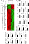
Figure 1
Figure 1. Microarray analysis of the effect of estrogen on global gene expression in mammary tissue during involution.
Mammary glands of mice at 48 h involution were treated with Ctrl(n = 3) or E2B in sesame oil(n = 3) for 24 h. Total mammary RNA was analyzed for E2B-induced gene expression using Affymetrix Mouse Gene 2.0 ST array.(a) Heat map of E2B-regulated genes with fold change ≥2.0 and p value < 0.05.(b) RT-PCR validation of a panel of 12 E2B regulated genes.(c) Known estrogen target genes Areg, Greb1 and Pgr were also upregulated after 24 h E2B treatment in involuting mice.(d) Estrogen-induced immune response gene expression except for Timp1 in involuting mammary does not occurs in the mammary tissue of nulliparous mice. Ctrl, n = 3; E2B, n = 3. All data are presented as Mean ± SEM. White and black bars represent Ctrl and E2B treatment, respectively. Statistical significance was evaluated by unpaired two-tailed Student’s t-tests; *p < 0.05, **p < 0.01, ***p < 0.001.
Figure 2
Figure 2. Estrogen-induced expression of immune response genes during mammary involution occurs mainly in leucocytes.
Mice at 24 h post-weaning were treated with Ctrl or E2B in sesame oil for 48 h. Mammary tissues were digested to obtain single cell suspension. CD45-positive leucocytes were separated from CD45− cells using Dynabeads®.(a) CD45-positive cell depletion efficiency was assessed using FACS.(b) RT-PCR analysis of the expression of various genes regulated by estrogen in total mammary population(TMP), CD45-depleted(CD45−) and CD45-positive(CD45+) cells. Results are presented as Mean ± SEM. n = 4 mice per group. For all graphs, white and black bars represent Ctrl- and E2B-treated group, respectively. Statistical significance was evaluated by unpaired two-tailed Student’s t-tests; *p < 0.05, **p < 0.01, ***p < 0.001. u.d., undetected.
Figure 3
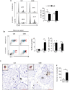
Figure 3. Estrogen promotes neutrophil recruitment during mammary tissue involution.
Mice at 24 h post-weaning or age-matched nulliparous mice were treated with Ctrl or E2B in sesame oil for 48 h. Mammary tissues were digested by collagenase to obtain single cell suspension. Mammary cells and blood samples were stained with various cell surface markers.(a) E2B treatment did not affect total CD45+ cell infiltration in involuting or nulliparous mammary tissue. Representative histograms were shown for CD45+ in Ctrl- and E2B-treated nulliparous and involuting mammary glands.(b) Estrogen treatment induced significant neutrophils and myeloid-derived monocytic cells infiltration to involuting but not to nulliparous mammary tissue.(Left) Representative dot plots of neutrophils(CD45+CD11b+Gr-1hi)(red boxes) and myeloid-derived monocytic cells(CD45+CD11b+ Gr1int)(blue boxes) from mammary glands CD45+ cell population.(Right) Bar graphs show the percentages of neutrophils and myeloid-derived monocytic cells in total cell population. For the data on mammary tissue in(a,b) from nulliparous mice, Ctrl n = 10, E2B n = 9; for the data of mammary tissue in(a,b) from mice undergoing involution, Ctrl n = 13, E2B n = 13.(c) Gr-1 IHC of Ctrl- or E2B-treated involuting mammary tissue. Scale bar = 50 μm.(d) Total number of infiltrated neutrophils in mammary tissue were counted in an area of 20 mm2 for each sample. n = 9 per group. White and black bars represent Ctrl and E2B treatment, respectively. Statistical significance was evaluated by unpaired two-tailed Student’s t-tests, **p < 0.01.
Figure 4
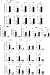
Figure 4. Estrogen targets neutrophils specifically to induce inflammatory gene signature.
(a) Mice at 24 h post-weaning were treated with Ctrl(n = 4) or E2B(n = 4) in sesame oil for 48 h. Neutrophils were isolated from mammary tissues using biotinylated anti-Gr-1 antibody coupled to Streptavidin Dynabeads® and total RNA from neutrophils were analyzed for gene expression by RT-PCR. The mRNA levels relative to 36B4 are expressed as Mean ± SEM.(b,c,d) Depletion of neutrophils abolishes estrogen-induced gene expression in involuting mammary glands. Mice at 24 h post-weaning were administered with isotype control IgG or Ly6G antibody followed by treatment with Ctrl or E2B in sesame oil for 48 h.(b) Ly6G antibody significantly reduced the percentages of neutrophils in blood(left) and mammary gland(right) by FACS analysis.(c,d) Neutrophil depletion abrogated E2B-induced expression of immune response genes but not the known estrogen target genes Greb1 and Pgr. Ctrl+IgG n = 4; E2B+IgG n = 4; Ctrl+Ly6G n = 3; E2B + Ly6G n = 3. White and black bars represent Ctrl and E2B treatment, respectively. Statistical significance was evaluated by unpaired two-tailed Student’s t-tests; *p < 0.05, **p < 0.01, ****p < 0.0001.
Figure 5
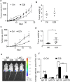
Figure 5. Estrogen-stimulated neutrophils are required for estrogen-induced mammary tumor growth in involuting mammary tissue.
(ad) Estrogen stimulated markedly ERα-negative 4T1 cell growth in involuting mammary gland.(a,b) 0.5 × 106 4T1-luc2 cells were injected into the ninth abdominal mammary gland at 24 h post-weaning. The mice were treated with Ctrl(n = 7) or E2B(n = 8) in sesame oil daily for the first 7 days after tumor injection. Volumes of palpable tumors were measured by digital calipers at 2–3 days intervals(a). Tumor weight at end point(day 37)(b). Ctrl n = 6;E2B n = 8.(c,d) Involuting mice were implanted with Ctrl or estradiol valerate(E2V) pellets at 24 h post-weaning. 1 × 106 4T1-luc2 cells were injected into the ninth abdominal mammary gland 24 h after the hormone pellet implantation. Volumes of palpable tumors were measured by digital calipers at 3 days intervals(c). Tumor weights at the time of sacrifice(day 15)(d). Ctrl n = 9; E2V n = 9.(e,f) Neutrophil depletion abolished E2B-induced tumor growth. Mice at 24 h post-weaning were given either isotype control IgG or Ly6G antibody followed by Ctrl or E2B treatment in sesame oil and were injected with 1 × 106 4T1-luc2 cells. Bioluminescence imaging of tumors was performed on day 4 and day 6 of tumor inoculation. Ctrl+IgG n = 4, E2B+IgG n = 4, Ctrl+Ly6G n = 4, E2B+Ly6G n = 4. Representative bioluminescence image of tumor-bearing mouse from each of the four groups on day 6 of tumor injection(e). Bioluminescent quantification of tumor growth(f). For figures(ad), statistical significance was evaluated by unpaired two-tailed Student’s t-tests; for figure(f), statistical significance was evaluated by one way ANOVA followed by post-hoc Tukey’s multiple comparisons test, **p < 0.01, ***p < 0.001.

References

    1. Lambe M. et al.. Transient increase in the risk of breast cancer after giving birth. N Engl J Med 331, 5–9, doi: 10.1056/NEJM199407073310102(1994). - DOI - PubMed
    1. Stein T., Salomonis N. & Gusterson B. A. Mammary gland involution as a multi-step process. J Mammary Gland Biol Neoplasia 12, 25–35, doi: 10.1007/s10911-007-9035-7(2007). - DOI - PubMed
    1. Lyons T. R. et al.. Postpartum mammary gland involution drives progression of ductal carcinoma in situ through collagen and COX-2. Nat Med 17, 1109–1115, doi: 10.1038/nm.2416(2011). - DOI - PMC - PubMed
    1. Lyons T. R. et al.. Cyclooxygenase-2-dependent lymphangiogenesis promotes nodal metastasis of postpartum breast cancer. J Clin Invest 124, 3901–3912, doi: 10.1172/JCI73777(2014). - DOI - PMC - PubMed
    1. Lecarpentier J. et al.. Breast Cancer Risk Associated with Estrogen Exposure and Truncating Mutation Location in BRCA1/2 Carriers. Cancer Epidemiol Biomarkers Prev 24, 698–707, doi: 10.1158/1055-9965.EPI-14-0884(2015). - DOI - PubMed

Publication types