. 2017 Apr 21:7:46627.
doi: 10.1038/srep46627.

Sensory system plasticity in a visually specialized, nocturnal spider

Affiliations

Sensory system plasticity in a visually specialized, nocturnal spider

Jay A Stafstrom et al. Sci Rep. .

Abstract

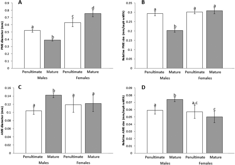
The interplay between an animal's environmental niche and its behavior can influence the evolutionary form and function of its sensory systems. While intraspecific variation in sensory systems has been documented across distant taxa, fewer studies have investigated how changes in behavior might relate to plasticity in sensory systems across developmental time. To investigate the relationships among behavior, peripheral sensory structures, and central processing regions in the brain, we take advantage of a dramatic within-species shift of behavior in a nocturnal, net-casting spider (Deinopis spinosa), where males cease visually-mediated foraging upon maturation. We compared eye diameters and brain region volumes across sex and life stage, the latter through micro-computed X-ray tomography. We show that mature males possess altered peripheral visual morphology when compared to their juvenile counterparts, as well as juvenile and mature females. Matching peripheral sensory structure modifications, we uncovered differences in relative investment in both lower-order and higher-order processing regions in the brain responsible for visual processing. Our study provides evidence for sensory system plasticity when individuals dramatically change behavior across life stages, uncovering new avenues of inquiry focusing on altered reliance of specific sensory information when entering a new behavioral niche.


Conflict of interest statement

The authors declare no competing financial interests.

Figures

Figure 1
Figure 1. Forward facing eyes of Deinopis spinosa (PME = posterior median eye, AME = anterior median eye).
Left and right columns represent photos from the same individual (A,C = male, B,D = female) across penultimate (A,B) and mature (C,D) life stages. All scale bars = 0.5 mm.
Figure 2
Figure 2
(A) Deinopis spinosa in foraging posture. While hanging in a support web (not detectable in this photograph), net-casting spiders will hold a specialized capture snare in their front legs, and actively entangle prey that walks beneath or flies above. (B,C) Internal anatomy within the cephalothorax of D. spinosa, (B) is a dorsal view, (C) is a lateral view. Protocerebrum (Proto) is red, ONPs are green, MBs are blue, AB is yellow. Anterior to the ONPs lies the optic nerve, which connects retinas from the eyes to the ONPs. The enlarged PMEs can be seen anterior to the optic nerve in (C).
Figure 3
Figure 3. Eye size comparisons across groups.
Significant differences are symbolized by differences in letters above respective bars. Absolute PME diameter (A) significantly differs across each group. While standardizing for body size using cephalothorax width measurements (B), mature males had relatively smaller PMEs than every other group, while no other group significantly differed from each other. Similarly, mature males had significantly larger absolute AME diameters (C) when compared to all other groups, while no other group was significantly different from each other. When standardizing for body size using cephalothorax measurements, mature males also had relatively larger AMEs than all other groups. Error bars signify one standard deviation above and below the average trait value.
Figure 4
Figure 4. Micro-CT scans of focal brain regions.
Dorsal views of segmented brain region outlines (AC) and reconstructed regions (DF). Focal regions depicted are the ONPs (A,D), the MBs (B,E), and the AB (C,F).
Figure 5
Figure 5. Neural structure comparisons across groups.
Significant differences are symbolized by differences in letters above respective bars. Relative ONP investment (A), standardized using total protocerebrum volume, is significantly lower in mature males when compared to every other group, while all other groups had similar relative investment in ONPs. Relative investment in the MBs (B), standardized using “central brain” total volume, was significantly greater in females overall, and lowest in mature males. Relative investment in the AB (C), also standardized using “central brain” volume, was highest in mature males and was significantly greater in mature males than in both penultimate and mature females. Error bars signify one standard deviation above and below the average trait value.
Figure 6
Figure 6. Relationships between relative investment in lower-order visual processing and higher-order integration centers across all focal groups (circles = male, square = female, white = penultimate, black = mature).
A positive relationship exists between relative ONP investment and relative investment in the MBs (A). In contrast, a negative relationship exists between relative ONP investment and relative AB investment (B). Both relationships are heavily influenced by mature male investment.

References

    1. Ronald K. L., Fernández-Juricic E. & Lucas J. R. Taking the sensory approach: How individual differences in sensory perception can influence mate choice. Anim. Behav. 84, 1283–1294 (2012).
    1. Wylie D. R., Gutierrez-Ibanez C. & Iwaniuk A. N. Integrating brain, behaviour and phylogeny to understand the evolution of sensory systems in birds. Front. Neurosci. 9, 1–17 (2015). - PMC - PubMed
    1. Poulson T. L. & White W. B. The cave environment. Science (80). 165, 971–981 (1969). - PubMed
    1. Niven J. E. & Laughlin S. B. Energy limitation as a selective pressure on the evolution of sensory systems. J. Exp. Biol. 211, 1792–804 (2008). - PubMed
    1. Soares D. & Niemiller M. L. Sensory adaptations of fishes to subterranean environments. Bioscience 63, 274–283 (2013).

Publication types

LinkOut - more resources