. 2018 Mar;128(3):875-884.
doi: 10.3171/2016.11.JNS16976. Epub 2017 Apr 21.

Noninvasive neuromodulation and thalamic mapping with low-intensity focused ultrasound

Affiliations

Noninvasive neuromodulation and thalamic mapping with low-intensity focused ultrasound

Robert F Dallapiazza et al. J Neurosurg. 2018 Mar.

Abstract

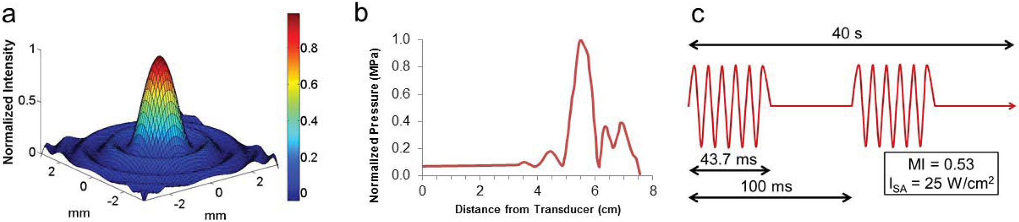
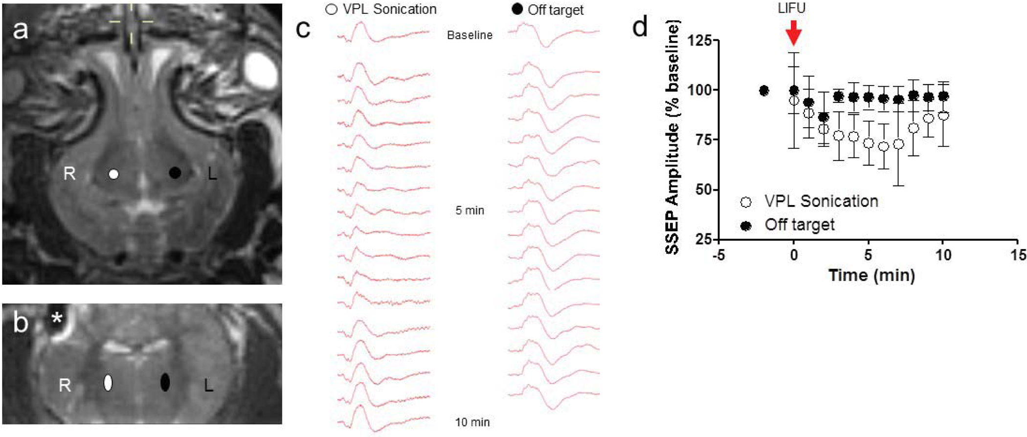
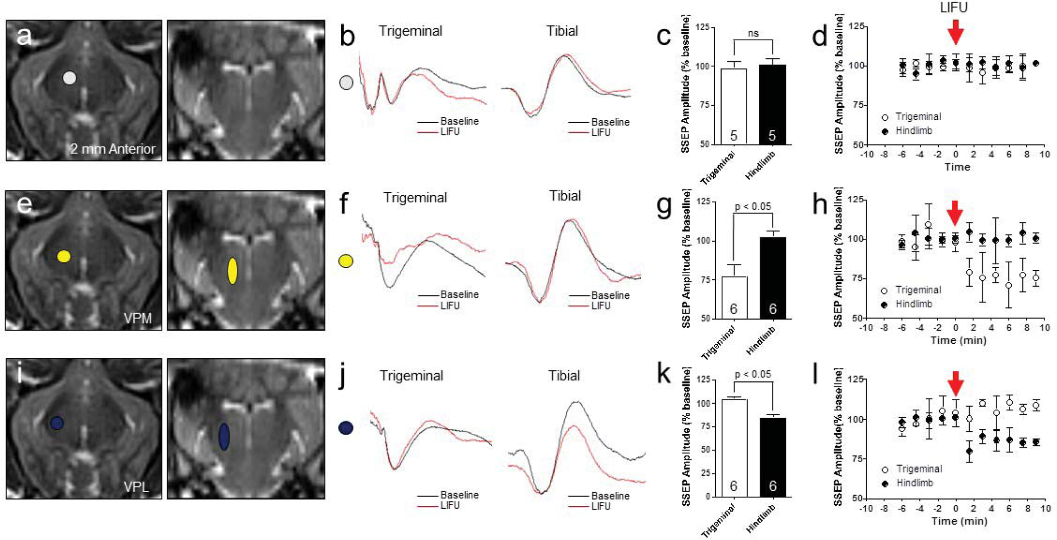
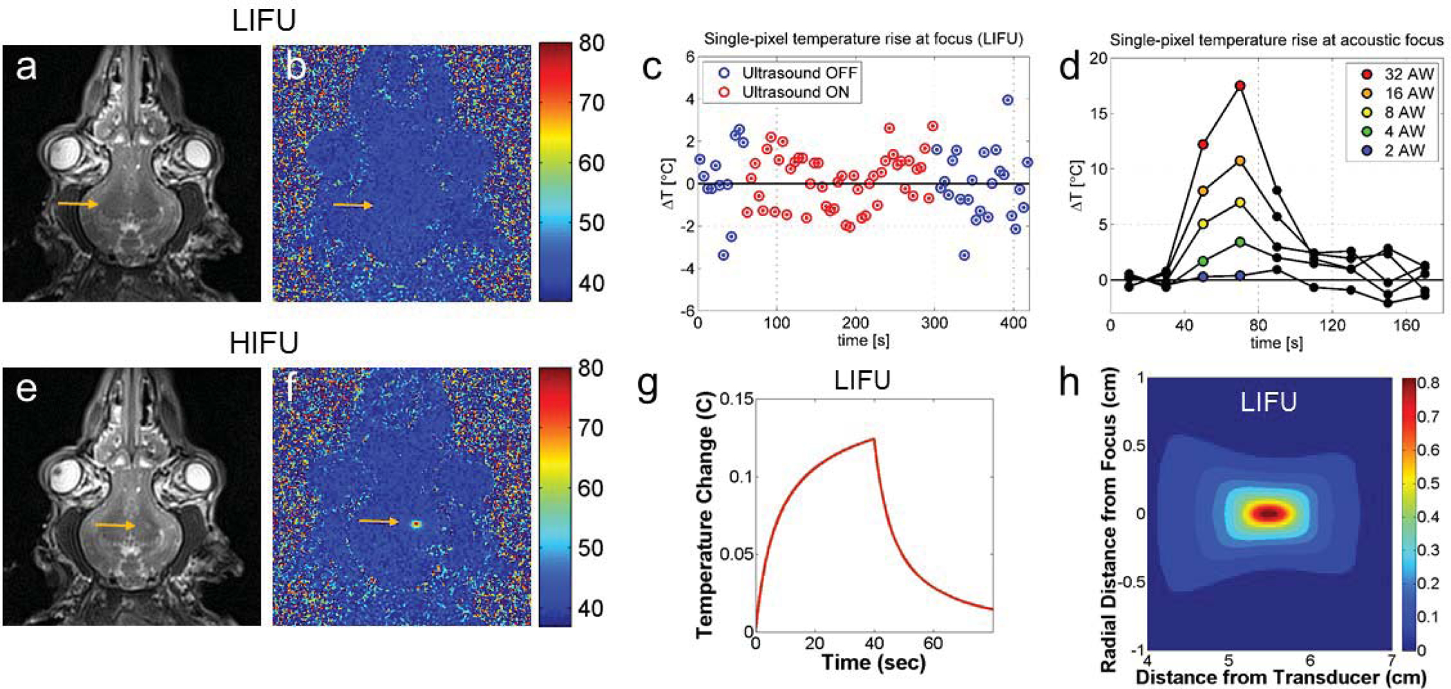
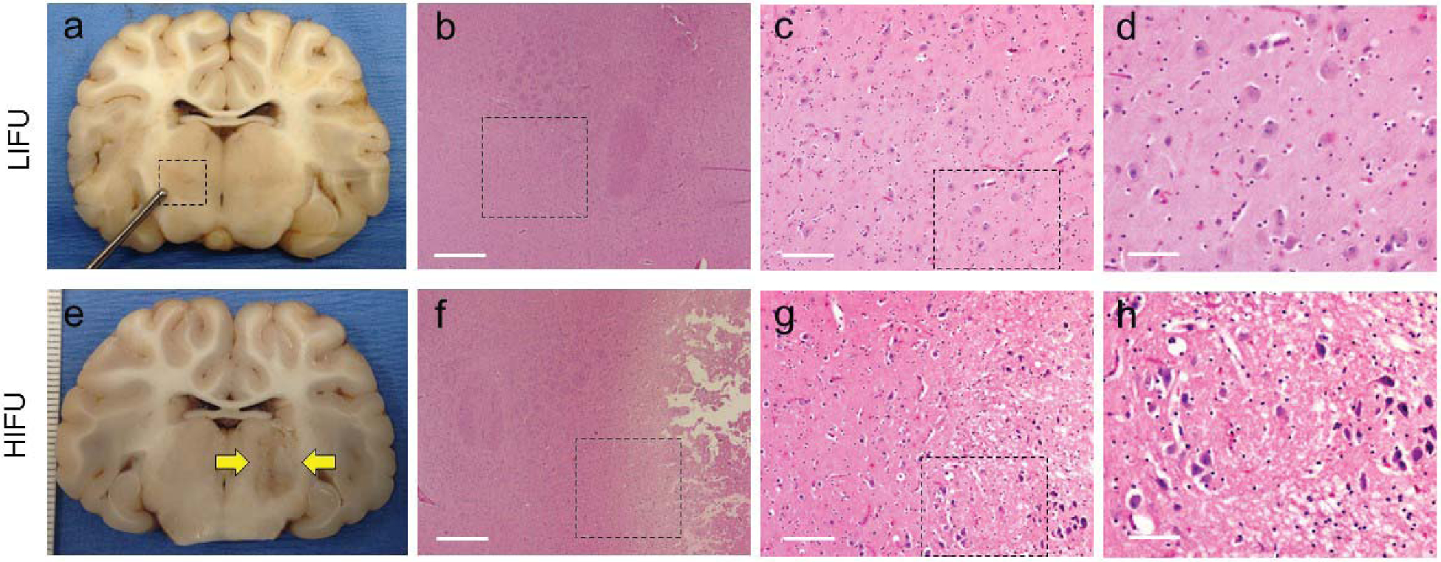
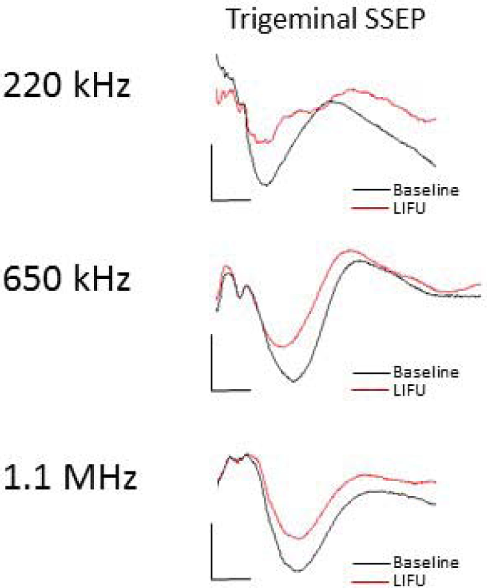
OBJECTIVE Ultrasound can be precisely focused through the intact human skull to target deep regions of the brain for stereotactic ablations. Acoustic energy at much lower intensities is capable of both exciting and inhibiting neural tissues without causing tissue heating or damage. The objective of this study was to demonstrate the effects of low-intensity focused ultrasound (LIFU) for neuromodulation and selective mapping in the thalamus of a large-brain animal. METHODS Ten Yorkshire swine ( Sus scrofa domesticus) were used in this study. In the first neuromodulation experiment, the lemniscal sensory thalamus was stereotactically targeted with LIFU, and somatosensory evoked potentials (SSEPs) were monitored. In a second mapping experiment, the ventromedial and ventroposterolateral sensory thalamic nuclei were alternately targeted with LIFU, while both trigeminal and tibial evoked SSEPs were recorded. Temperature at the acoustic focus was assessed using MR thermography. At the end of the experiments, all tissues were assessed histologically for damage. RESULTS LIFU targeted to the ventroposterolateral thalamic nucleus suppressed SSEP amplitude to 71.6% ± 11.4% (mean ± SD) compared with baseline recordings. Second, we found a similar degree of inhibition with a high spatial resolution (∼ 2 mm) since adjacent thalamic nuclei could be selectively inhibited. The ventromedial thalamic nucleus could be inhibited without affecting the ventrolateral nucleus. During MR thermography imaging, there was no observed tissue heating during LIFU sonications and no histological evidence of tissue damage. CONCLUSIONS These results suggest that LIFU can be safely used to modulate neuronal circuits in the central nervous system and that noninvasive brain mapping with focused ultrasound may be feasible in humans.

Keywords: FUS = focused ultrasound; H & E = hematoxylin and eosin; HIFU = high-intensity focused ultrasound; ISA = spatial average intensity; LFB = Luxol fast blue; LIFU = low-intensity focused ultrasound; PRFS = proton resonance frequency shift; SSEP = somatosensory evoked potential; VPL = ventroposterolateral thalamic nucleus; VPM = ventroposteromedial thalamic nucleus; brain mapping; functional neurosurgery; low intensity focused ultrasound; neuromodulation; noninvasive; somatosensory evoked potentials; thalamus.


Figures

Figure 1.
Figure 1.
Ultrasound parameters and characterization. (a) The single element 1.14 MHz focused transducer has a narrow 1 × 1 mm focus in the lateral dimension (b) and a 5 mm focus in the axial direction, with a focal length of 5.6 cm. (c) Neuromodulation treatments were performed with a 43. 7% duty cycle over 40 seconds, with an ISA of 25–30 W/cm2 maintained across all three (1.14 MHz, 650 kHz, 220 kHz) ultrasound systems used.
Figure 2.
Figure 2.
Thalamic neuromodulation with LIFU. (a, b) Axial and coronal T2-weighted MRI demonstrating the right VPL target (white), control left VPL target (black), and artifact from the cortical electrode(*). (c) Representative median nerve SSEP at baseline, 5 minutes and 10 minutes following right VPL LIFU sonication and control, ‘off’ target sonications. Note the suppression in SSEPs at 5 minutes after sonication, with a return to baseline by 10 minutes. (d) LIFU delivered to the right VPL decreased left median nerve SSEP to 71.6 ± 11.4 % (mean ± SD) compared to baseline recordings (N = 9 sonications, 4 animals). SSEPs recovered to 87.2 ± 15.4 % baseline values within 10 minutes. Control sonications in the left VPL had no lasting effects on left median nerve SSEP, which were 96.3 ± 7.4 % of baseline values (N = 6 sonications, 4 animals).
Figure 3.
Figure 3.
Noninvasive thalamic nucleus mapping with LIFU. (a, e, i) Thalamic nucleus targeting for control (2 mm anterior to VPM, white shape), VPM (yellow shapes), and VPL (blue shapes) with LIFU. (b, f, j) Representative trigeminal- and tibial-SSEPs at baseline (black) and post-sonication (red). (c, d) Nontarget, control LIFU did not alter either the trigeminal-evoked or tibial-evoked SSEP compared to baseline recordings (trigeminal, 98.2 ± 4.5 % and tibial, 100.6 ± 4.4 %; p = 0.40 and 0.42, respectively), (N = 5 sonications, 4 animals). The acoustic focus was 2 mm anterior to the VPM target (g, h) Right VPM LIFU decreased trigeminal-SSEP to 76.9 ± 7.5 % of baseline values (N = 6 sonications, 4 animals; p = 0.0002), but tibial-SSEP were not significantly changed at 102.0 ± 4.3 % compared to baseline values (N = 6 sonications, 4 animals; p = 0.19). (k, l) Right VPL LIFU had no significant effect on trigeminal-SSEP (103.9 ± 3.3 %, p > 0.05), but tibial-SSEP decreased significantly to 83.9 ± 4.3 % compared to baseline values (N = 6 sonications, 4 animals, p = 4.2 × 10⁻⁶).
Figure 4.
Figure 4.
LIFU temperature monitoring. (a) T2-weighted image post LIFU showing no change in signal at the acoustic focus. (b) MR proton shift resonance image showing no change in temperature during LIFU. (c) Temperature changes at the acoustic focus measured every 5 seconds during a LIFU sonication with a 240-second duration showing a 0.1 ± 0.3 °C average temperature change. Blue points correspond to baseline MR images, and red points correspond to MR images acquired during sonication. (d) Temperature changes with increasing sonication power confirm that there is no temperature increase until the acoustic power is increased by a factor of 16 (from 0.25 Wto 4 W). (e) T2*-weighted image after HIFU showing small hyperintensity at the acoustic focus. (f) MR proton shift resonance image confirms temperature elevations during HIFU. (g) Modeled temperature rise at the acoustic focus during LIFU sonication predicted by the HIFU Simulator V1.2. The predicted peak temperature, 0.13 °C, occurs at sonication termination. (d) Modeled thermal map showing predicted peak spatial temperature rise during LIFU with a 1 × 1 × 3 mm focal heating <0.05 °C.
Figure 5.
Figure 5.
Gross and histological analysis of thalamic LIFU and HIFU. (a-d) LIFU sonications demonstrating no evidence of histological damage. H&E taken at 4×, 10×, and 40×. Black boxes represent the area depicted in the following panel. (e-h) HIFU sonication showing well-circumscribed lesion in the ventrolateral thalamus. Microscopic analysis shows ischemic neurons along the periphery and edema extending into the white matter tracts. Black boxes represent the area depicted in the following panel.
Figure 6.
Figure 6.
Thalamic LIFU neuromodulation occurs across ultrasound frequencies. Representative trigeminal-evoked potentials with LIFU focused at the VPM nucleus demonstrating neuromodulation using three separate ultrasound transducers with parent frequencies 220 kHz, 710 kHz, and 1.1 MHz.

References

    1. Adrianov OS, Vykhodtseva NI, Fokin VF, Uranova NA, Avirom VM: [Reversible functional shutdown of the optic tract on exposure to focused ultrasound]. Biull Eksp Biol Med 97:760–762, 1984 - PubMed
    1. Chang WS, Jung HH, Kweon EJ, Zadicario E, Rachmilevitch I, Chang JW: Unilateral magnetic resonance guided focused ultrasound thalamotomy for essential tremor: practices and clinicoradiological outcomes. J Neurol Neurosurg Psychiatry 86:257–264, 2015 - PubMed
    1. Clement GT, Hynynen K: A non-invasive method for focusing ultrasound through the human skull. Physics in Medicine & Biology 47:1219–1236, 2002 - PubMed
    1. Clement GT, Sun J, Giesecke T, Hynynen K: A hemisphere array for non-invasive ultrasound brain therapy and surgery. Phys Med Biol 45:3707–3719, 2000 - PubMed
    1. Clement GT, White J, Hynynen K: Investigation of a large-area phased array for focused ultrasound surgery through the skull. Phys Med Biol 45:1071–1083., 2000 - PubMed

Publication types