. 2017 May;9(5):431-439.
doi: 10.1038/nchem.2644. Epub 2016 Nov 14.

Engineering genetic circuit interactions within and between synthetic minimal cells

Affiliations

Engineering genetic circuit interactions within and between synthetic minimal cells

Katarzyna P Adamala et al. Nat Chem. 2017 May.

Abstract

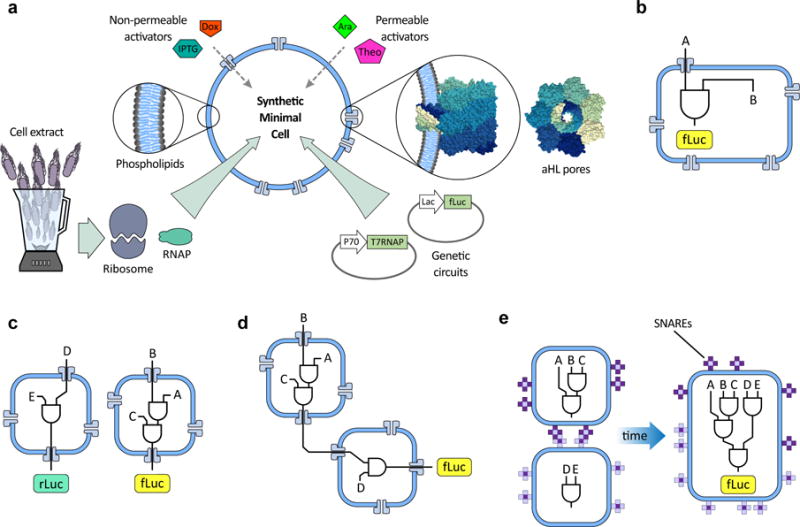
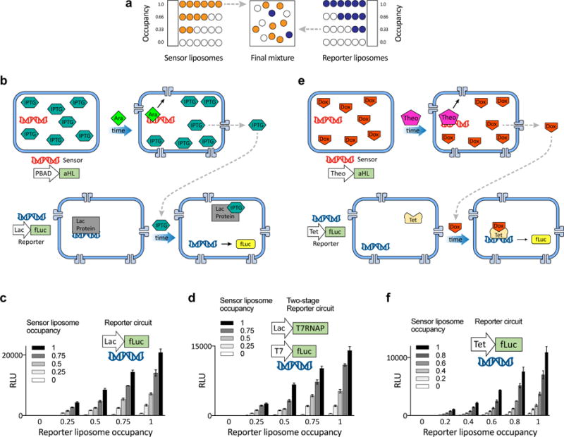
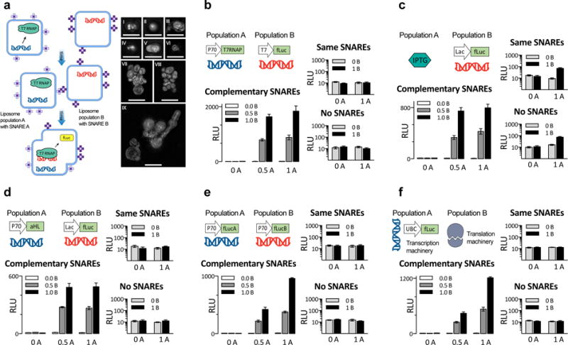
Genetic circuits and reaction cascades are of great importance for synthetic biology, biochemistry and bioengineering. An open question is how to maximize the modularity of their design to enable the integration of different reaction networks and to optimize their scalability and flexibility. One option is encapsulation within liposomes, which enables chemical reactions to proceed in well-isolated environments. Here we adapt liposome encapsulation to enable the modular, controlled compartmentalization of genetic circuits and cascades. We demonstrate that it is possible to engineer genetic circuit-containing synthetic minimal cells (synells) to contain multiple-part genetic cascades, and that these cascades can be controlled by external signals as well as inter-liposomal communication without crosstalk. We also show that liposomes that contain different cascades can be fused in a controlled way so that the products of incompatible reactions can be brought together. Synells thus enable a more modular creation of synthetic biology cascades, an essential step towards their ultimate programmability.


Figures

Fig. 1
Fig. 1. An overview of genetic circuit interactions within and between synthetic minimal cells (synells)
a. Synthetic minimal cells (synells) are semipermeable compartments made from a phospholipid bilayer membrane and various contents. The membrane can display a variety of proteins, including channel-forming proteins such as alpha-hemolysin (aHL). The phospholipid membranes of synells are permeable to molecules such as theophylline (Theo) and arabinose (Ara), and are permeable to others like β-D-1-thiogalactopyranoside (IPTG) and doxycycline (Dox) when aHL channels are present; these molecules can be used for triggering activity within synells. Synells can encapsulate cell lysates with transcriptional and/or translational activity, as well as DNA vectors encoding genes. In this paper, we demonstrate four novel competencies of synells that, together, can be used to create complex, modular genetic circuits. b. We show that synells can contain genetic circuits in which all the components and operations take place within the same liposome. c. We show that two genetic circuits can work independently in separate liposome populations. d. We show that genetic circuits within two different liposome populations can interact. e. We show that genetic circuits can run in parallel in separate compartmentalized reactions; if those reactions are encapsulated by liposomes carrying fusogenic peptides such as SNAREs, the reaction products can be joined together in a hierarchical fashion.
Fig. 2
Fig. 2. Molecular confinement of multicomponent genetic cascades
a. Images of liposomes expressing GFP. Sub-panels I–IV: structured illumination microscopy (SIM) images of representative liposomes expressing GFP and membrane-labeled with rhodamine. Every SIM image (panels labeled I, II, III, IV) represents a separate liposome; all liposomes were imaged on the same day and all liposomes came from the same sample, prepared 24h before imaging. All SIM images in this figure are at the same scale; the large scale bars in panels I and II are 1 μm, the small scale bars in panels III and IV are 200 nm. Sub-panels V–VI: widefield epifluorescent images of liposomes expressing GFP. The liposomes for this imaging sample were extruded through a 2 μm filter and dialyzed with a 1 μm membrane; panel V shows sample after 6 h incubation and panel VI shows an aliquot of the same sample after 24 h incubation. The scale bars on panels V and VI are 10 μm. b–d. Fraction of synells expressing GFP and split GFP, measured by flow cytometry (for control flow cytometry experiments, see Fig. S2). b. Bulk expression of GFP and fluorescence measured on the sample prior to the flow cytometry experiments. c. Analysis of samples expressing GFP; 68.4% of liposomes produced measurable green signal. d. Analysis of samples expressing split GFP; 61.8% of liposomes produced measurable green signal.
Fig 3
Fig 3. Comparison of single- and multi-component genetic circuits
a–c. Genetic cascades involving one-, two-, or three-part luciferase protein assemblies. Expressed under doxycycline-inducible Tet promoters were whole firefly luciferase (fLuc) (a), the two halves (here denoted fLucA and fLucB) of split fLuc bearing split inteins and mutually binding coiled coils (b), and two halves (here denoted fLucC and fLucD) of split fLuc bearing split inteins and coiled coils that bind to a third common template (denoted “scaffold”) (c). d–f. Effects of dilution on fLuc expression in liposomes vs. bulk solution, for the fLuc assemblies described in a–c (see Fig. S6 for experiments under the control of a constitutive P70 promoter). Dotted lines throughout this figure are visual guides, not fits. RLU, relative light units. g–i. End-point expression of luciferase measured at the 3 h time point, for 7 different concentrations of doxycycline (Dox). See Fig. S7 for corresponding 1 h end-point expression data, and Figs. S8 – S10 for the same reactions in bulk solution. j–l. Comparison of liposomal vs. bulk solution expression of luciferase, at 2 different time points and for 10 ng/mL of Dox. The 2 plasmids in k and 3 plasmids in l were mixed at equimolar ratios, with total DNA concentration held constant. Error bars indicate S. E. M. n=4 replicates.
Fig. 4
Fig. 4. Insulation of genetic circuits operating in parallel liposome populations
a. Schematic of liposome populations designed to contain similar genetic components but to respond differently to the same environmental concentration of the non-membrane-permeable small molecule activator doxycycline (Dox), by expressing different amounts of the alpha-hemolysin channel protein (aHL). These liposomes contain a measured amount of the plasmid for constitutively expressed aHL, and of a plasmid driving either firefly luciferase (fLuc) or Renilla luciferase (rLuc) from the Tet inducible promoter (the luciferase plasmids were always held at the same concentration). Throughout this figure, the two populations were incubated together in the solution containing Dox and harvested after 6 h (see Figs. S11 and S12 for rLuc and fLuc expression as a function of aHL plasmid concentration, after 2 h and 6 h, respectively). b. Each liposome contains either 0.1 nM or 5 nM of the aHL plasmid. c. Luciferase expression in symmetrical populations, where the amount of aHL DNA is the same across the two populations; the amount of fLuc and rLuc expression is graphed with respect to aHL plasmid concentration and to each other. d–e. Luciferase expression in asymmetrical populations. d. Luciferase expression when Renilla liposomes have a constant aHL plasmid concentration (0.1 nM) but the concentration of that plasmid is varied in the firefly liposomes. Expression of rLuc and fLuc are graphed against the plasmid concentration in firefly liposomes and against each other. e. Luciferase expression as in d, but with constant aHL plasmid concentration in firefly liposomes and variable concentration in Renilla liposomes. Error bars indicate S. E. M. n=4 replicates.
Fig. 5
Fig. 5. Communication between genetic circuits operating in multiple liposome populations
a. Scheme for mixing two populations of liposomes at different ratios of their components while maintaining a constant lipid concentration of 10 mM (the same scheme was used throughout this figure and Fig. 6). Each population contains the same amount of liposomes, but the liposome occupancy can vary between 0 (all liposomes are empty) and 1 (the maximum fraction of the liposomes contain reagents). b–d. Externally activated two-part circuits, with bacterial TX/TL. b. Scheme of interacting populations, denoted sensor and reporter. Sensor liposomes contain the alpha-hemolysin gene and are filled with IPTG; reporter liposomes contain machinery for firefly luciferase expression. During activation, arabinose (Ara) diffuses through the sensor liposome membrane and induces aHL expression, which releases IPTG, which induces fLuc expression in the reporter. c. Expression of fLuc for varying ratios of occupancy (as in a), for the sensor and reporter liposomes with indicated contents. This panel represents the time point 6h (for complete time series see Fig. S14). For this circuit without arabinose see Fig. S15. d. Expression of fLuc for a circuit in which the reporter liposomes contain DNA for a multicomponent genetic cascade, as indicated. This panel represents the 6h time point (for complete time series, see Fig. S16. For this circuit without arabinose, see Fig. S17). e–f. Externally activated two-part circuits, containing both bacterial and mammalian TX/TL components. e. Sensor vesicles contain the Theo-triggered aHL gene and Dox; reporter liposomes contain constitutively expressed aHL and Tet, and Dox/Tet-driven fLuc. During activation, Theo diffuses through the membrane of the activator liposomes and induces aHL expression, which creates pores that release Dox from the activator. Dox induces fLuc expression in the reporter liposomes. f. Expression of fLuc, for varying ratios of sensor and reporter liposomes (this panel represents 6h time point; for complete time series see Fig. S18. For this circuit without Theo, see Fig. S19). Error bars indicate S. E. M. n=4 replicates.
Fig. 6
Fig. 6. Fusion of complementary genetic circuits
a. General scheme for SNARE-mediated liposome fusion. We created two populations of liposomes, A and B, decorated with complementary SNARE protein mimics in their outer leaflet. The images to the right, in sub-panels I through IX, are maximum-intensity projections of structured illumination microscopy (SIM) z-stacks of liposomes membrane-labeled with rhodamine, bearing complementary SNARE pairs, and fused for 4 hours. All images from panels I through IX represent separate fields of view. Scale bars, 5 μm. All liposomes in this figure, except f, contained bacterial TX/TL components. b–f. Five different instantiations of the liposome fusion concept, exploring several ways to distribute genetic circuits across fusable liposomes, with two different populations of liposomes at three occupancy levels for each case. b. Mixing of constitutively expressed T7 RNA polymerase with firefly Luciferase under T7 promoter. c. Mixing of a non-membrane-permeable small molecule activator IPTG with its inducible promoter, driving fLuc production. d. Mixing of a constitutively expressed membrane channel with an inducible promoter driving fLuc production, in the background of the small molecule that induces the promoter (IPTG). e. Mixing liposomes with genes encoding split protein. f. Mixing liposomes containing mammalian transcription (HeLa) and translation (HeLa) system, producing fLuc. For all five systems in b–f., experiments on the large graph are with one of a matching pair of SNARE on each population, the top of the two small panels is both liposomes with the same SNARE, and the bottom one neither population had any SNAREs. In both small graphs of b–f, the y-axis is in logarithmic scale to show the near-zero values for non-fusing liposomes. Switching which liposome contained which SNARE had no effect on the results (Fig. S25), whereas the absence of SNARE proteins or the presence of identical SNAREs on both populations hindered fusions (small graphs on b–f). Error bars indicate S. E. M. n=4 replicates.

References

    1. Carlson ED, Gan R, Hodgman CE, Jewett MC. Cell-free protein synthesis: Applications come of age. Biotechnol Adv. 2012;30:1185–1194. - PMC - PubMed
    1. Smith MT, Wilding KM, Hunt JM, Bennett AM, Bundy BC. The emerging age of cell-free synthetic biology. FEBS Lett. 2014;588:2755–2761. - PubMed
    1. Hodgman CE, Jewett MC. Cell-free synthetic biology: Thinking outside the cell. Metab Eng. 2012;14:261–269. - PMC - PubMed
    1. Miller D, Gulbis J. Engineering Protocells: Prospects for Self-Assembly and Nanoscale Production-Lines. Life. 2015;5:1019–1053. - PMC - PubMed
    1. Shimizu Y, Kuruma Y, Ying BW, Umekage S, Ueda T. Cell-free translation systems for protein engineering. FEBS J. 2006;273:4133–4140. - PubMed

Publication types