

Age-related reduction in the expression of FOXO transcription factors and correlations with intervertebral disc degeneration
- PMID: 28430387
- PMCID: PMC5650945
- DOI: 10.1002/jor.23583
Age-related reduction in the expression of FOXO transcription factors and correlations with intervertebral disc degeneration
Abstract
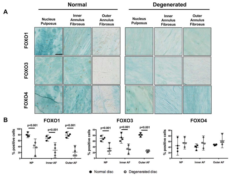
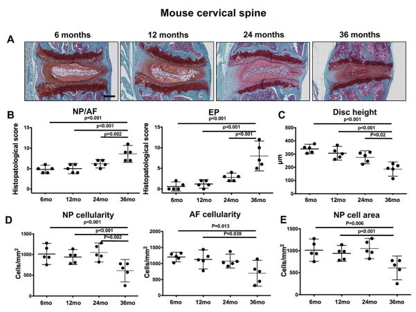
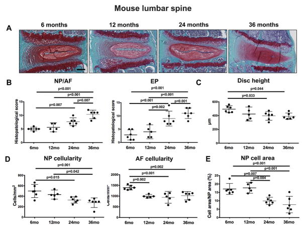
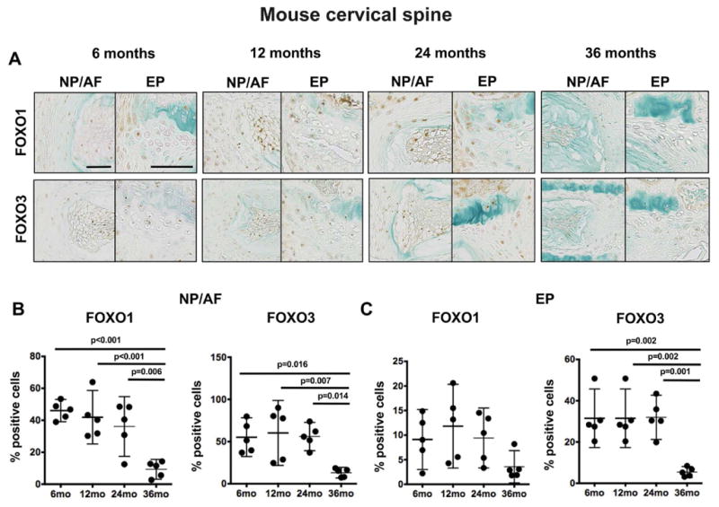
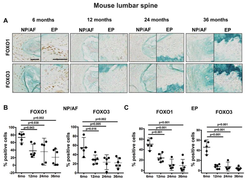
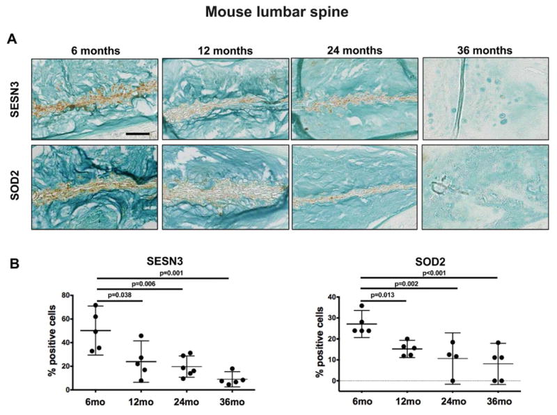
Aging is a main risk factor for intervertebral disc (IVD) degeneration, the main cause of low back pain. FOXO transcription factors are important regulators of tissue homeostasis and longevity. Here, we determined the expression pattern of FOXO in healthy and degenerated human IVD and the associations with IVD degeneration during mouse aging. FOXO expression was assessed by immunohistochemistry in normal and degenerated human IVD samples and in cervical and lumbar IVD from 6-, 12-, 24-, and 36-month-old C57BL/6J mice. Mouse spines were graded for key histological features of disc degeneration in all the time points and expression of two key FOXO downstream targets, sestrin 3 (SESN3) and superoxide dismutase (SOD2), was assessed by immunohistochemistry. Histological analysis revealed that FOXO proteins are expressed in all compartments of human and mouse IVD. Expression of FOXO1 and FOXO3, but not FOXO4, was significantly deceased in human degenerated discs. In mice, degenerative changes in the lumbar spine were seen at 24 and 36 months of age whereas cervical IVD showed increased histopathological scores at 36 months. FOXO expression was significantly reduced in lumbar IVD at 12-, 24-, and 36-month-old mice and in cervical IVD at 36-month-old mice when compared with the 6-month-old group. The reduction of FOXO expression in lumbar IVD was concomitant with a decrease in the expression of SESN3 and SOD2. These findings suggest that reduced FOXO expression occurs in lumbar IVD during aging and precedes the major histopathological changes associated with lumbar IVD degeneration. © 2017 Orthopaedic Research Society. Published by Wiley Periodicals, Inc. J Orthop Res 35:2682-2691, 2017.
Keywords: FOXO; aging; intervertebral disc.
© 2017 Orthopaedic Research Society. Published by Wiley Periodicals, Inc.
Figures
Similar articles
-
FOXO are required for intervertebral disk homeostasis during aging and their deficiency promotes disk degeneration.Aging Cell. 2018 Oct;17(5):e12800. doi: 10.1111/acel.12800. Epub 2018 Jul 2. Aging Cell. 2018. PMID: 29963746 Free PMC article.
-
Microcalcification of lumbar spine intervertebral discs and facet joints is associated with cartilage degeneration, but differs in prevalence and its relation to age.J Orthop Res. 2017 Dec;35(12):2692-2699. doi: 10.1002/jor.23591. Epub 2017 May 18. J Orthop Res. 2017. PMID: 28467655
-
Ectopic expression of Smurf2 and acceleration of age-related intervertebral disc degeneration in a mouse model.J Neurosurg Spine. 2017 Jul;27(1):116-126. doi: 10.3171/2016.11.SPINE16901. Epub 2017 Apr 7. J Neurosurg Spine. 2017. PMID: 28387615
-
Cell death in intervertebral disc degeneration.Apoptosis. 2013 Jul;18(7):777-85. doi: 10.1007/s10495-013-0839-1. Apoptosis. 2013. PMID: 23512131 Review.
-
The molecular basis of intervertebral disc degeneration.Spine J. 2013 Mar;13(3):318-30. doi: 10.1016/j.spinee.2012.12.003. Epub 2013 Feb 8. Spine J. 2013. PMID: 23537454 Review.
Cited by
-
Scientific literature landscape analysis of researches on oxidative stress in intervertebral disc degeneration in web of science.Front Mol Biosci. 2022 Aug 11;9:989627. doi: 10.3389/fmolb.2022.989627. eCollection 2022. Front Mol Biosci. 2022. PMID: 36032668 Free PMC article.
-
PHLPP1 deficiency protects against age-related intervertebral disc degeneration.JOR Spine. 2022 Sep 27;5(4):e1224. doi: 10.1002/jsp2.1224. eCollection 2022 Dec. JOR Spine. 2022. PMID: 36601379 Free PMC article.
-
p300 arrests intervertebral disc degeneration by regulating the FOXO3/Sirt1/Wnt/β-catenin axis.Aging Cell. 2022 Aug;21(8):e13677. doi: 10.1111/acel.13677. Epub 2022 Jul 30. Aging Cell. 2022. PMID: 35907249 Free PMC article.
-
Epigenetic changes during aging and their reprogramming potential.Crit Rev Biochem Mol Biol. 2019 Feb;54(1):61-83. doi: 10.1080/10409238.2019.1570075. Epub 2019 Mar 1. Crit Rev Biochem Mol Biol. 2019. PMID: 30822165 Free PMC article. Review.
-
Reciprocal Regulation of TRPS1 and miR-221 in Intervertebral Disc Cells.Cells. 2019 Sep 28;8(10):1170. doi: 10.3390/cells8101170. Cells. 2019. PMID: 31569377 Free PMC article.
References
-
- Dagenais S, Caro J, Haldeman S. A systematic review of low back pain cost of illness studies in the United States and internationally. Spine J. 2008;8(1):8–20. - PubMed
-
- Manchikanti L, Singh V, Falco FJ, Benyamin RM, Hirsch JA. Epidemiology of low back pain in adults. Neuromodulation. 2014;17(Suppl 2):3–10. - PubMed
-
- The Department of Economic and Social Affairs of the United Nations. World Population Prospects. The 2015 Revision. http://esa.un.org/unpd/wpp/Publications/FIles/Key_FIndings_WPP_2015.pdf2015.
-
- Luoma K, Riihimaki H, Luukkonen R, Raininko R, Viikari-Juntura E, Lamminen A. Low back pain in relation to lumbar disc degeneration. Spine (Phila Pa 1976) 2000;25(4):487–92. - PubMed
-
- Deyo RA, Weinstein JN. Low back pain. N Engl J Med. 2001;344(5):363–70. - PubMed
Publication types
MeSH terms
Substances
Grants and funding
LinkOut - more resources
Full Text Sources
Other Literature Sources
Medical
Research Materials
Miscellaneous
