. 2017 May 30;8(22):36423-36437.
doi: 10.18632/oncotarget.16855.

Parkin regulates translesion DNA synthesis in response to UV radiation

Affiliations

Parkin regulates translesion DNA synthesis in response to UV radiation

Xuefei Zhu et al. Oncotarget. .

Abstract

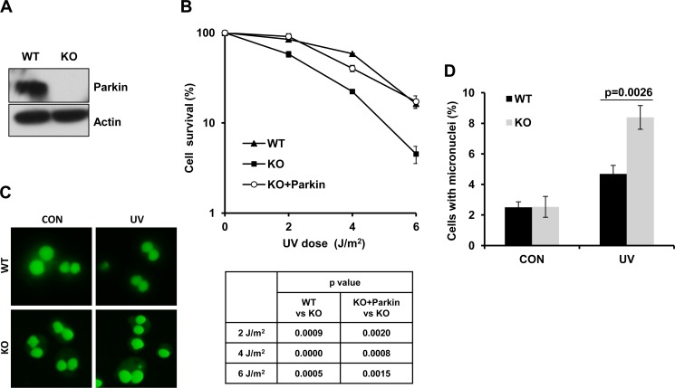
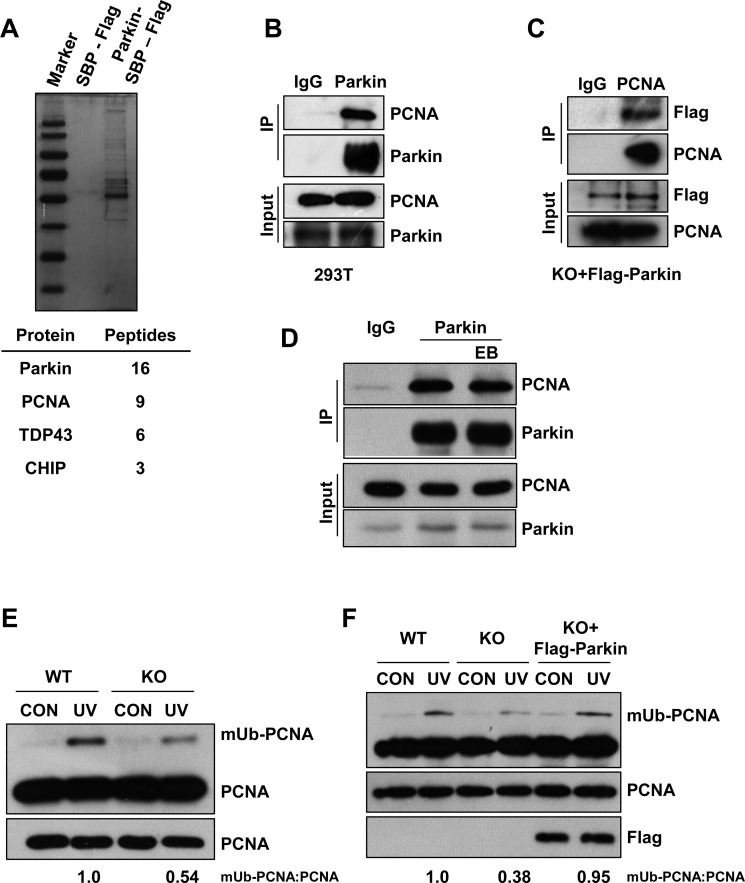
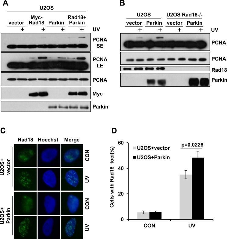
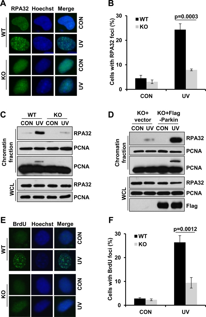
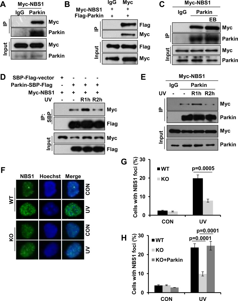
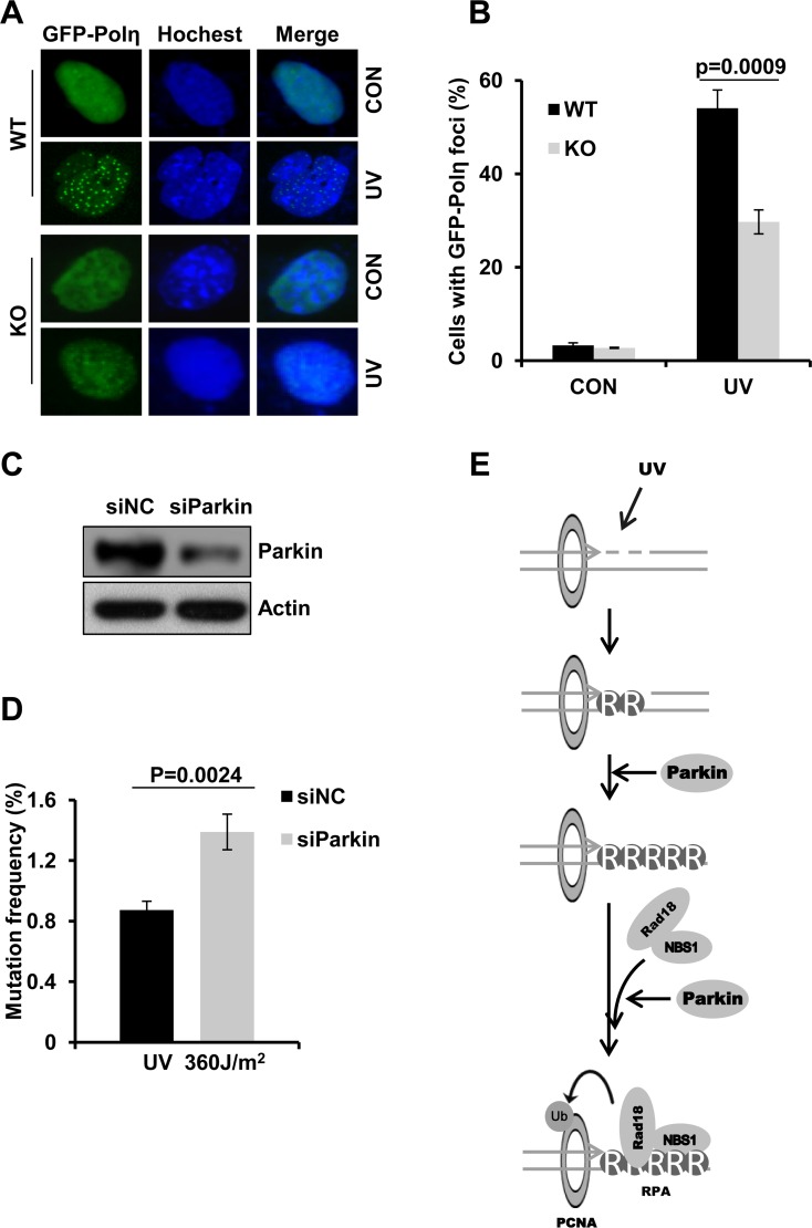
Deficiency of Parkin is a major cause of early-onset Parkinson's disease (PD). Notably, PD patients also exhibit a significantly higher risk in melanoma and other skin tumors, while the mechanism remains largely unknown. In this study, we show that depletion of Parkin causes compromised cell viability and genome stability after ultraviolet (UV) radiation. We demonstrate that Parkin promotes efficient Rad18-dependent proliferating cell nuclear antigen (PCNA) monoubiquitination by facilitating the formation of Replication protein A (RPA)-coated ssDNA upon UV radiation. Furthermore, Parkin is found to physically interact with NBS1 (Nijmegen breakage syndrome 1), and to be required for optimal recruitment of NBS1 and DNA polymerase eta (Polη) to UV-induced damage sites. Consequently, depletion of Parkin leads to increased UV-induced mutagenesis. These findings unveil an important role of Parkin in protecting genome stability through positively regulating translesion DNA synthesis (TLS) upon UV damage, providing a novel mechanistic link between Parkin deficiency and predisposition to skin cancers in PD patients.

Keywords: Parkin; Parkinson’s disease; melanoma; translesion DNA synthesis; ultraviolet radiation.


Conflict of interest statement

CONFLICTS OF INTEREST

The authors declare no conflicts of interests.

Figures

Figure 1
Figure 1. Parkin-null cells are hypersensitive to UV radiation
(A) Expression of Parkin in WT and Parkin−/− cells were examined by western-blot with an anti-Parkin antibody. β-actin: loading control. (B) Viability of cells after exposure to UV radiation. WT, Parkin−/−, and Parkin−/− cells complemented with Parkin were irradiated with the indicated doses of UV, and further incubated in complete medium for 10 days, then cell colonies were determined. Cell viability was expressed as a percentage of mock-treated cells. Error bars represent SD, t-test, n = 3. (CD) WT and Parkin−/− cells were irradiated with 2 J/m2 UV, and further incubated in complete medium containing 6 μg/ml CB for 48 h. Then percentage of cells with micronuclei was determined. (C) Representative images of control and UV-irradiated WT and Parkin−/− cells in micronucleus test. (D) Quantification of the percentage of cells with micronuclei. Error bars represent SD. t-test, n = 3.
Figure 2
Figure 2. Parkin promotes PCNA monoubiquitination after exposure to UV radiation
(A) HEK293T cells ectopically expressing empty vector or Parkin-SBP-Flag were used for tandem affinity purification as described in “Materials and Methods”. The final eluates were resolved by SDS-PAGE and revealed by silver staining. Representative proteins identified by mass spectrum were shown below. (B) HEK293T cells were lysed and incubated with protein A/G agarose conjugated with normal rabbit IgG or anti-Parkin antibody, the immunoprecipitates were then examined via western blot with antibodies against PCNA and Parkin. (C) Parkin−/− cells stably expressing Flag-Parkin were lysed and incubated with protein A/G agarose conjugated with normal mouse IgG or anti-PCNA antibody, the immunoprecipitates were then examined via western blot with antibodies against PCNA and Flag. (D) HEK293T cells were lysed and incubated with protein A/G agarose conjugated with normal rabbit IgG or anti-Parkin antibody for immunoprecipitation in the presence of EB or not, followed by blotting with the indicated antibodies. (E) WT and Parkin−/− cells were irradiated with 15 J/m2 UV and collected 3 h later. The levels of mUb-PCNA were examined by western blot with an anti-PCNA antibody. (F) WT, Parkin−/−, and Parkin−/− cells complemented with Flag-Parkin were treated and examined as in (E).
Figure 3
Figure 3. Parkin promotes Rad18 dependent PCNA monoubiquitination
(A) U2OS cells ectopically expressing Myc-Rad18, Parkin or Myc-Rad18 and Parkin were irradiated with 15 J/m2 UV and collected 3 h later. The levels of mUb-PCNA were examined by western blot with an anti-PCNA antibody. SE, short exposure; LE, long exposure. (B) WT and Rad18−/− U2OS cells transfected with empty vector or Parkin were treated and examined as in (A). (CD) U2OS cells transfected with empty vector or Parkin were irradiated with 15 J/m2 UV and recovered for 3 h. Cells were pre-extracted with 0.5% Triton for 10 min and then fixed, immunostained with anti-Rad18 antibody and Hoechst-stained. (C) Representative images of cells stained with Hoechst or antibody against Rad18 after UV radiation. (D) Quantification of the percentage of cells with more than 20 Rad18 foci. Error bars represent SD. t-test, n = 3.
Figure 4
Figure 4. Parkin facilitates ssDNA generation and efficient RPA recruitment in response to UV radiation
(AB) WT and Parkin−/− cells were irradiated with 15 J/m2 UV and further incubated for 2 h. Cells were pre-extracted with 0.5% Triton for 10 min and then fixed, immunostained with anti-RPA32 antibody and Hoechst-stained. (A) Representative images of cells stained with Hoechst or antibody against RPA32 after UV radiation. (B) Quantification of the percentage of cells with more than 20 RPA32 foci. Error bars represent SD. t-test, n = 3. (C) WT and Parkin−/− cells were irradiated with 15 J/m2 UV and further incubated for 2 h. The chromatin fraction were harvested and separated by SDS-PAGE. The levels of RPA32 were detected by western blot with an anti-RPA32 antibody. The levels of RPA32 and PCNA in whole-cell lysates (WCL) were also detected. (D) Parkin−/− cells complemented with Flag-Parkin or empty vector were treated and examined as in (C). (EF) WT and Parkin−/− cells were incubated with 10 μM BrdU for 48 h before irradiated with 15 J/m2 UV, and further incubated for 2 h. Cells were pre-extracted with 0.5% Triton and immunostained with anti-BrdU antibody and Hoechst-stained. (E) Representative images of cells stained with Hoechst or antibody against BrdU after UV radiation. (F) Quantification of the percentage of cells with more than 20 BrdU foci. Error bars represent SD. t-test, n = 3.
Figure 5
Figure 5. Parkin interacts with NBS1 and regulates its redistribution after exposure to UV radiation
(AB) Interaction between Parkin and NBS1. HEK293T cells ectopically expressing Myc-NBS1 (A) or Myc-NBS1 and Flag-Parkin (B) were lysed and incubated with protein A/G agarose conjugated with either anti-Parkin or anti-Myc antibody. The immunoprecipitated products were blotted with the indicated antibodies. (C) HEK293T cells ectopically expressing Myc-NBS1 were lysed and incubated with protein A/G agarose conjugated with normal rabbit IgG or anti-Parkin antibody for immunoprecipitation in the presence of EB or not, followed by blotting with the indicated antibodies. (DE) Dynamic association between Parkin and NBS1 upon UV radiation. (D) HEK293T cells transfected with Myc-NBS1 and Parkin-SBP-Flag or empty vector were irradiated with 15 J/m2 UV and recovered for either 1 h (R1h) or 2 h (R2h). Cell lysates were immunoprecipitated with streptavidin-conjugated sepharose followed by blotting with the indicated antibodies. (E) HEK293T cells ectopically expressing Myc-NBS1 were treated as above. Cell lysates were immunoprecipitated with protein A/G agarose conjugated with normal rabbit IgG or anti-Parkin antibody followed by blotting with the indicated antibodies. (F-H) NBS1 foci formation after UV radiation. WT, Parkin−/−, and Parkin−/− cells complemented with Parkin were irradiated with 15 J/m2 UV and further incubated for 2 h. Cells were pre-extracted with 0.5% Triton for 10 min and then immunostained with anti-NBS1 antibody and Hoechst-stained. (F) Representative images of cells stained with Hoechst or antibody against NBS1 after UV radiation. (GH) Quantification of the percentage of cells with more than 20 NBS1 foci. Error bars represent SD. t-test, n = 3.
Figure 6
Figure 6. Parkin deficiency attenuates Polη recruitment to damage sites and leads to higher mutation frequency
(AB) Parkin deficiency was detrimental for recruitment of Polη to UV-induced damage sites. WT or Parkin−/− cells transfected with GFP-Polη were irradiated with 15 J/m2 UV and further incubated for 8 h. Then cells were fixed and the proportion of cells with GFP-Polη foci was determined. (A) Representative images of Polη foci formation in WT and Parkin−/− cells. (B) Quantification of the percentage of cells with more than 30 GFP-Polη foci. Error bars represent SD. t-test, n = 3. (CD) Parkin deficiency led to higher mutation frequency. (C) 293T cells were transfected with siParkin or siNC. 72 h later, the cells were harvested and the levels of Parkin were detected by western blot. β-actin: loading control. (D) The Parkin-depleted 293T cells were transfected with pSP189 plasmid which was pre-irradiated with 360 J/m2 UV, and replicated for 72 h. The pSP189 plasmid was then extracted and DpnI digested followed by transformation into MBM7070 bacteria strain for mutation frequency analysis. Error bars represent SD. t-test, n = 3. (E) Model for Parkin's function in regulating TLS.

Similar articles

Cited by

References

    1. Kitada T, Asakawa S, Hattori N, Matsumine H, Yamamura Y, Minoshima S, Yokochi M, Mizuno Y, Shimizu N. Mutations in the parkin gene cause autosomal recessive juvenile parkinsonism. Nature. 1998;392:605–8. doi: 10.1038/33416. - DOI - PubMed
    1. Lucking CB, Durr A, Bonifati V, Vaughan J, De Michele G, Gasser T, Harhangi BS, Meco G, Denefle P, Wood NW, Agid Y, Brice A, French Parkinson's Disease Genetics Study G et al. Association between early-onset Parkinson's disease and mutations in the parkin gene. N Engl J Med. 2000;342:1560–7. doi: 10.1056/NEJM200005253422103. - DOI - PubMed
    1. Shimura H, Hattori N, Kubo S, Mizuno Y, Asakawa S, Minoshima S, Shimizu N, Iwai K, Chiba T, Tanaka K, Suzuki T. Familial Parkinson disease gene product, parkin, is a ubiquitin-protein ligase. Nat Genet. 2000;25:302–5. doi: 10.1038/77060. - DOI - PubMed
    1. Riley BE, Lougheed JC, Callaway K, Velasquez M, Brecht E, Nguyen L, Shaler T, Walker D, Yang Y, Regnstrom K, Diep L, Zhang Z, Chiou S, et al. Structure and function of Parkin E3 ubiquitin ligase reveals aspects of RING and HECT ligases. Nat Commun. 2013;4:1982. doi: 10.1038/ncomms2982. - DOI - PMC - PubMed
    1. Dawson TM, Dawson VL. The role of parkin in familial and sporadic Parkinson's disease. Mov Disord. 2010;25:S32–9. doi: 10.1002/mds.22798. - DOI - PMC - PubMed

MeSH terms