Review
. 2017 May 16;8(20):33972-33989.
doi: 10.18632/oncotarget.15687.

The regulation of β-catenin activity and function in cancer: therapeutic opportunities

Affiliations
Review

The regulation of β-catenin activity and function in cancer: therapeutic opportunities

Shuang Shang et al. Oncotarget. .

Abstract

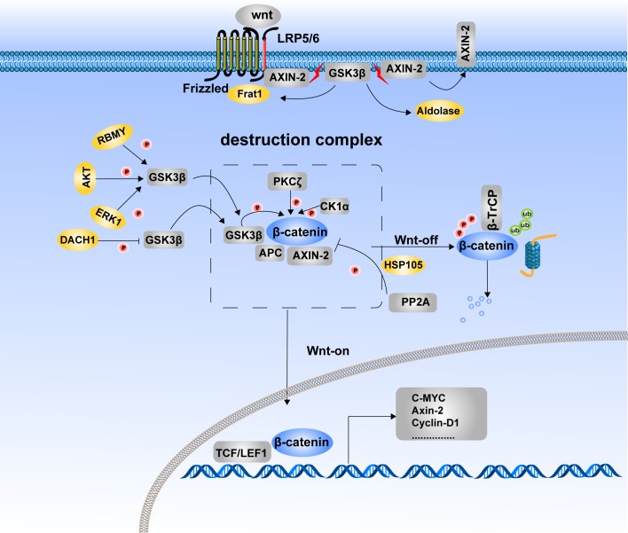
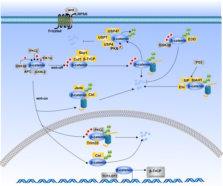
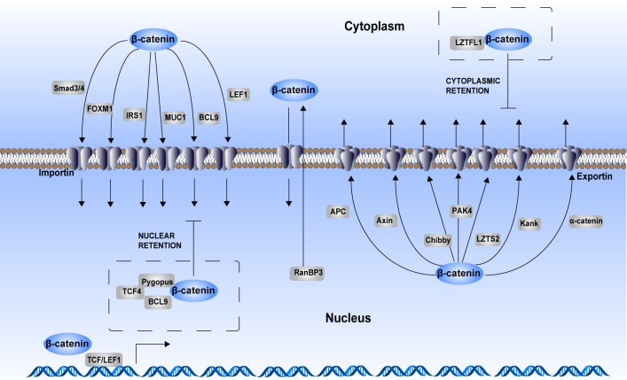
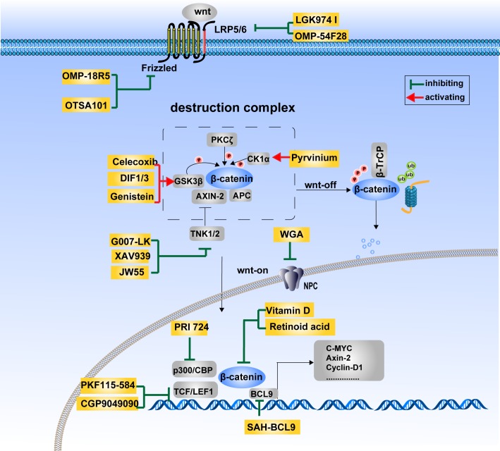
Wnt/β-catenin signaling is an evolutionarily conserved and versatile pathway that is known to be involved in embryonic development, tissue homeostasis and a wide variety of human diseases. Aberrant activation of this pathway gives rise to the accumulation of β-catenin in the nucleus and promotes the transcription of many oncogenes such as c-Myc and CyclinD-1. As a result, it contributes to carcinogenesis and tumor progression of several cancers, including colon cancer, hepatocellular carcinoma, pancreatic cancer, lung cancer and ovarian cancer. β-Catenin is a pivotal component of the Wnt signaling pathway and it is tightly regulated at three hierarchical levels: protein stability, subcellular localization and transcriptional activity. Uncovering the regulatory mechanisms of β-catenin will provide new insights into the pathogenesis of cancer and other diseases, as well as new therapeutic strategies against these diseases. In this review we dissect the concrete regulatory mechanisms of β-catenin from three aspects mentioned above. Then we focus on the role of β-catenin in cancer initiation, progression, dormancy, immunity and cancer stem cell maintenance. At last, we summarize the recent progress in the development of agents for the pharmacological modulation of β-catenin activity in cancer therapy.

Keywords: Wnt signaling; cancer therapy; protein stability; subcellular localization; transcriptional regulation.


Conflict of interest statement

CONFLICTS OF INTEREST

We declare that we have no conflicts of interest.

Figures

Figure 1
Figure 1. Phosphorylation of β-catenin and its degradation
The phosphorylated status of β-catenin is determined for its stability. Destruction complex formation is the requested step for β-catenin phosphorylation. In the Wnt-off state, β-catenin was recruited to the destruction complex and induced phosphorylation and subsequent degradation. In the Wnt-on state, β-catenin translocates into the nucleus due to the disassembly of the destruction complex. LRP5/6, FRAT1 and aldolase inhibit β-catenin degradation by suppressing assembly of the destruction complex, which provides a platform for β-catenin phosphorylation. Kinases and phosphatases jointly take part in the balance control of β-catenin between phosphorylation and dephosphorylation. GSK3β, CK1α and PKCζ are protein kinases that catalyze β-catenin phosphorylation and its subsequent degradation. PP2A is the phosphatase that induces dephosphorylation of β-catenin and inhibits its degradation. RBMY, AKT, ERK1, and DACT1 indirectly regulate the phosphorylation and degradation of β-catenin by modulating the Ser9 phosphorylation of GSK3β to inactivate its kinase activity.
Figure 2
Figure 2. Ubiquitination of β-catenin and its degradation
The degradation of β-catenin mainly undergoes through Ubiquitin Proteosome System (UPS). Different ubiquitin E3 ligases in the cytoplasm and nucleus regulate the ubiquitination of β-catenin. Ubiquitination of β-catenin can be occurred either in the Wnt-off state or in the Wnt-on state. Together with Cul1 and Skp1, β-TrCP mediates the ubiquitination of β-catenin in the Wnt-off state. While Jade1, c-Cbl and Trim33 induce ubiquitination of β-catenin in the Wnt-on state. Additionally, SIAH1 along with SIP and Ebi, ubiquitinate β-catenin in response to p53 activition. Phosphrylation state is not always a necessary condition for β-catenin ubiquitination. β-TrCP and Trim33-induced ubiquitination of β-catenin is phosphorylation dependent, while ubiquitination of β-catenin by Jade-1, c-Cbl and SIAH1 are phosphorylation independent. In contrast to afore mentioned E3 ligases, EDD ubiquitinates β-catenin with an inhibitory effects on its degradation. Several deubiquitinases such as USP4 and USP7 can deubiquitinate β-catenin and interrupt its degradation process.
Figure 3
Figure 3. Regulation of β-catenin cytoplasmic–nuclear trafficking
As a consequence of aberrant activation of Wnt signaling, β-catenin translocates into the nucleus to act as a transcriptional activator. Although β-catenin itself does not have a nuclear localization sequence (NLS), its armadillo repeats mediate its nuclear translocation process. Many chaperones participate in the regulation of β-catenin cytoplasmic–nuclear trafficking. Smad3/4, FOXM1, IRS1, MUC1, BCL9 and LEF1 are chaperones that assist nuclear translocation of β-catenin in an importin-dependent manner. APC, AXIN, Chibby, PAK4, LZTS2, Kank, and α-catenin are chaperones that assist with β-catenin cytoplasmic translocation in an exportin-dependent manner. RanBP3 also enhances β-catenin nuclear export, while this process is independently of exportin. In addition, some proteins form complex with β-catenin to maintain its cytoplasmic or nuclear retention. LZTFL1 binds with β-catenin to sequester it in the cytoplasm. TCF4–Pygopus–BCL9 complex helps enhancing β-catenin nuclear retention.
Figure 4
Figure 4. Therapeutics targeting the Wnt/β-catenin signaling pathway
The Wnt/β-catenin signaling pathway is at the center of carcinogenesis and tumor progression. Therapeutics targeting this pathway is of great potential in the development of anti-cancer drugs. The agents currently being investigated mainly targets Wnt ligands and receptors (LGK974 I, OMP-54F28, OMP-18R5, OTSA101), the β-catenin destruction complex (Celecoxib, DIF1/3, Genistein, Pyrvinium, G007-LK, XAV939, JW55), β-catenin cytoplasmic–nuclear translocation (WGA) and β-catenin transcriptional complexes (PRI 724, Vitamin D, Retinoid acid, PKF115-584, CGP9049090, SAH-BCL9).

Similar articles

Cited by

References

    1. van Ooyen A, Nusse R. Structure and nucleotide sequence of the putative mammary oncogene int-1; proviral insertions leave the protein-encoding domain intact. Cell. 1984;39:233–240. - PubMed
    1. Behrens J, von Kries JP, Kühl M, Bruhn L, Wedlich D, Grosschedl R, Birchmeier W. Functional interaction of beta-catenin with the transcription factor LEF-1. Nature. 1996;382:638–642. - PubMed
    1. Peifer M, Pai LM, Casey M. Phosphorylation of the Drosophila adherens junction protein Armadillo: roles for wingless signal and zeste-white 3 kinase. Dev Biol. 1994;166:543–556. - PubMed
    1. Rubinfeld B, Albert I, Porfiri E, Fiol C, Munemitsu S, Polakis P. Binding of GSK3beta to the APC-beta-catenin complex and regulation of complex assembly. Science. 1996;272:1023–1026. - PubMed
    1. Yost C, Torres M, Miller JR, Huang E, Kimelman D, Moon RT. The axis-inducing activity, stability, and subcellular distribution of beta-catenin is regulated in Xenopus embryos by glycogen synthase kinase 3. Genes Dev. 1996;10:1443–1454. - PubMed