. 2017 Jul 25;8(30):48972-48982.
doi: 10.18632/oncotarget.16903.

P2X7 receptor and NLRP3 inflammasome activation in head and neck cancer

Affiliations

P2X7 receptor and NLRP3 inflammasome activation in head and neck cancer

Ju Young Bae et al. Oncotarget. .

Abstract

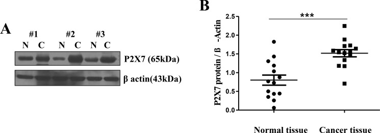
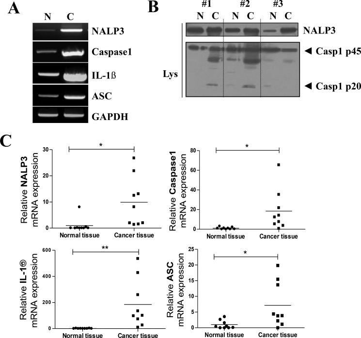
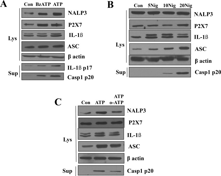
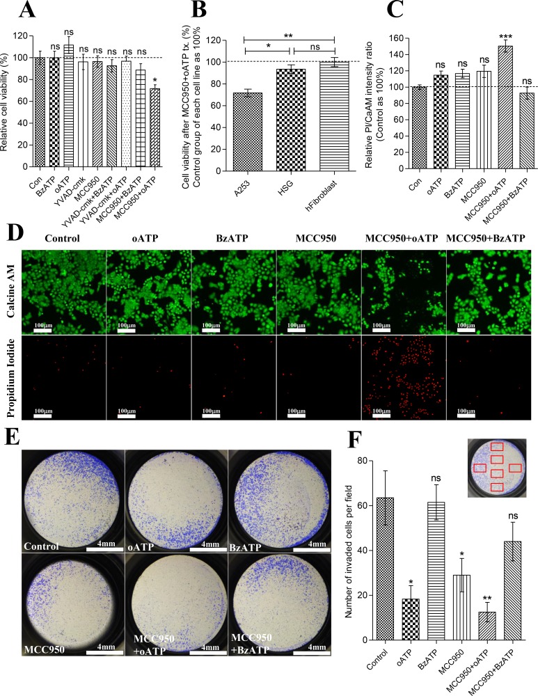
In this study, we investigated purinergic receptor P2X7 and NACHT, LRR and PYD domains-containing protein 3 (NLRP3) inflammasome expressions, and their role in head and neck cancer. We found upregulation of purinergic receptor P2X7 and all NLRP3 inflammasome components in biopsied head and neck squamous cell carcinoma tissues. Similarly, the expression of purinergic receptor P2X7, apoptosis-associated speck-like protein containing CARD, and pro-form caspase 1 in A253 cells derived from epidermoid carcinoma were highly upregulated in comparison to normal Human Salivary Gland cell line. Active caspase-1 and its final product, active interleukin-1β, both increased in primed A253 cells stimulated with purinergic receptor P2X7 agonists, while this elevated NLRP3 inflammasome activity was suppressed by purinergic receptor P2X7 antagonists. However, we observed none of these effects in Human Salivary Gland cells. Inhibition of both NLRP3 inflammasome and purinergic receptor P2X7 led to the significant cell death of primed A253 cells, but had no effect on the viability of primed HSG cells or the primary cultured human fibroblast cells. Furthermore, inhibition of either purinergic receptor P2X7 or NLRP3 inflammasome decreased invasiveness of A253, and this effect became more evident when both purinergic receptor P2X7 and NLRP3 inflammasome were simultaneously blocked. Therefore, it is concluded that the purinergic receptor P2X7 and the activation of NLRP3 inflammasome play important roles in the survival and invasiveness of head and neck squamous cell carcinoma in humans.

Keywords: A253 cells; NLRP3 inflammasome; head and neck squamous cell carcinoma; invasiveness; purinergic receptor P2X7.


Conflict of interest statement

CONFLICTS OF INTEREST

The authors declare no conflicts of interest.

Figures

Figure 1
Figure 1. Over-expression of P2X7R in oral cancer tissue
(A) Excised HNSCC and normal tissues were collected from three different patients to assess the expression level of P2X7R by Western blot (Lanes 1, 3, 5: normal tissues; lanes 2, 4, 6: oral cancer tissues). (B) The expression levels of P2X7R were significantly higher in the HNSCC patient group than in the control group (***P < 0.001, n = 14).
Figure 2
Figure 2. Over-expression of inflammasome components in oral cancer tissue
(A) mRNA expression of the inflammasome components NLRP3, caspase-1, and ASC and the inflammatory cytokines IL-1β. GAPDH is shown as a loading control. (B) A representative Western blot of three normal human tissue (N) and three HNSCC samples shows the protein expressions of NLRP3 protein, pro-caspase-1 (45 kDa), and activated caspase-1 p20 (20 kDa). (C) qPCR data collected from nine HNSCC patients' tumor and normal tissues. Raw data from normal tissues of each group was averaged and set as 1, thus fold change of each individual sample based on this value was plotted (n = 9, *P < 0.05, **P < 0.01).
Figure 3
Figure 3. Upregulation of P2X7R and NLRP3 inflammasome components in A253 cells
(A) The mRNA expression of NLRP3 inflammasome components in A253 and HSG cells. THP-1 cells, a macrophage cell line, were used as a positive control. Cells were treated with LPS for priming. RT-PCR analysis shows detectable levels of mRNA coding for members of the NLRP3 inflammasome complex. Results shown are representative of 3–4 individual experiments. (B) Expression of NLRP3 inflammasome in HSG and A253 cells at 0, 2, 6, and 19 h after stimulation with 1 μg/ml LPS. (C) NLRP3 inflammasome signaling is dispensable for ATP dose-dependent caspase-1 activation induced by LPS in A253 cells.
Figure 4
Figure 4. Role of P2X7R in the activation of NLRP3 inflammasome
(A) LPS-primed A253 cells were stimulated with 0.3 mM 2,3-O-(4-benzoyl-benzoyl)ATP (BzATP) for 3 h except for ATP that was added 30 min before the end of the experiment. Treatment with 5 mM ATP or 0.3 mM BzATP induced the release of caspase-1. (B) A253 cells were stimulated for 6 h with the indicated amounts (per ml) of nigericin. The range of nigericin concentrations was from 5 μM to 20 μM. Results shown are representative of 3–4 individual experiments (C) ATP or oxATP induced the release of caspase-1 in LPS-primed A253 cells.
Figure 5
Figure 5. Activity of P2X7R and NLRP3 inflammasome is closely related to the survival and invasiveness of A253 cells
(A) CCK-8 cell viability assay of LPS-primed A253 cells treated with oATP (1 μM), BzATP (0.3 mM), caspase-1 inhibitor (Ac-YVAD-cmk; 30 μg/ml), MCC950 (10 μM), or in combination of one another for 24hrs. Absorbance values at 450nm of control group were averaged and set as 100%. Data are expressed as the mean ± SEM of n = 6 (*P = < 0.05). (B) CCK-8 cell viability assay of LPS-primed A253, HSG, and primary cultured human fibroblast cells treated with combination of oATP (1 mM)+MCC950(10 μM). Absorbance values at 450 nm of each cell line's control group were averaged and set as 100%. Data are expressed as the mean ± SEM of n = 6 (*P = < 0.05, **P < 0.01). (C) Relative fluorescence intensity ratio of Propodium Iodide/Calcein AM. LPS-primed A253 cells were treated with oATP (1 mM), BzATP (0.3 mM), MCC950 (10 μM), or in combination of one another for 24 hrs. FL intensity ratios of PI/CaAM of control group were averaged and set as 100%. Data are expressed as the mean ± SEM of n = 6 (*P = < 0.05, **P < 0.01). (D) Epifluorescence image of PI/CaAM. LPS-primed A253 cells were treated with oATP (1 mM), BzATP (0.3 mM), MCC950 (10 μM), or in combination of one another for 24 hrs. FL images of PI channel were equally enhanced by increasing 40% brightness and 40% contrast. FL images of CaAM are unmodified images. (E) Matrigel invasion assay result. LPS-primed A253 cells were treated with oATP (1 mM), BzATP (0.3 mM), MCC950 (10 μM), or in combination for 24 hrs and trypsinized. 50000 viable cells were reseeded in each insert of the invasion chamber. 24 hrs later, invaded cells were fixed and stained with 1% crystal violet, and images of entire well were captured. 3 independent experiments were carried out. (F) Number of invaded cells per 20× magnified field was manually counted. Six different area of insert were selected and captured by Inverted microscope with 20X magnification. Selected areas are described in the graph as red squares. Data are expressed as the mean ± SEM of n = 6 (*P = < 0.05, **P < 0.01).

References

    1. Surprenant A, Rassendren F, Kawashima E, North RA, Buell G. The cytolytic P2Z receptor for extracellular ATP identified as a P2X receptor (P2X7) Science. 1996;272:735–738. - PubMed
    1. Adinolfi E, Raffaghello L, Giuliani AL, Cavazzini L, Capece M, Chiozzi P, Bianchi G, Kroemer G, Pistoia V, Di Virgilio F. Expression of P2X7 receptor increases in vivo tumor growth. Cancer Res. 2012;72:2957–2969. - PubMed
    1. Deli T, Varga N, Adam A, Kenessey I, Raso E, Puskas LG, Tovari J, Fodor J, Feher M, Szigeti GP, Csernoch L, Timar J. Functional genomics of calcium channels in human melanoma cells. Int J Cancer. 2007;121:55–65. - PubMed
    1. Raffaghello L, Chiozzi P, Falzoni S, Di Virgilio F, Pistoia V. The P2X7 receptor sustains the growth of human neuroblastoma cells through a substance P-dependent mechanism. Cancer Res. 2006;66:907–914. - PubMed
    1. Zhang XJ, Zheng GG, Ma XT, Yang YH, Li G, Rao Q, Nie K, Wu KF. Expression of P2X7 in human hematopoietic cell lines and leukemia patients. Leuk Res. 2004;28:1313–1322. - PubMed

MeSH terms

Substances