. 2017 Jun 1;140(6):1768-1783.
doi: 10.1093/brain/awx074.

Repurposed drugs targeting eIF2α-P-mediated translational repression prevent neurodegeneration in mice

Affiliations

Repurposed drugs targeting eIF2α-P-mediated translational repression prevent neurodegeneration in mice

Mark Halliday et al. Brain. .

Abstract

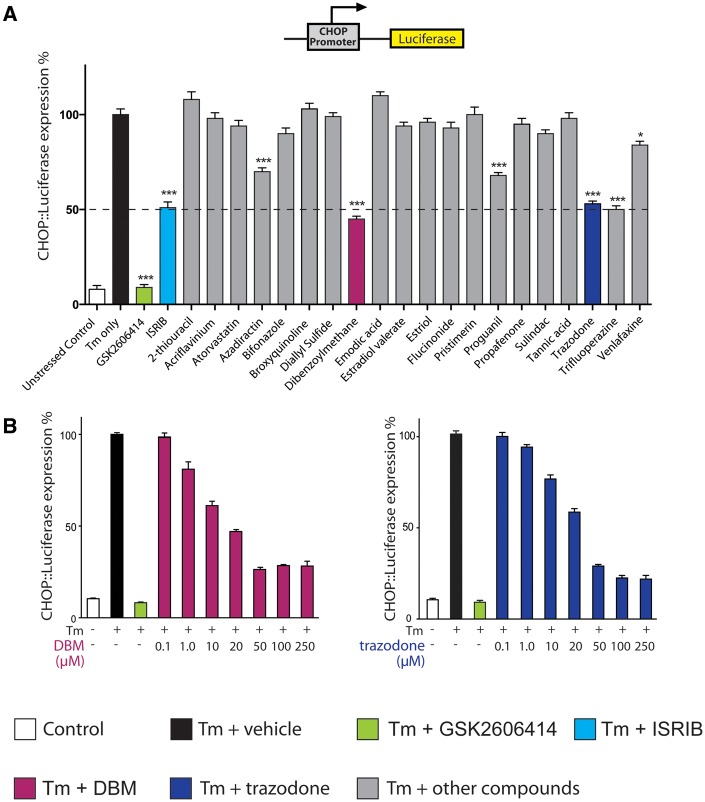
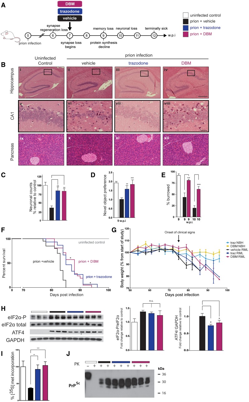
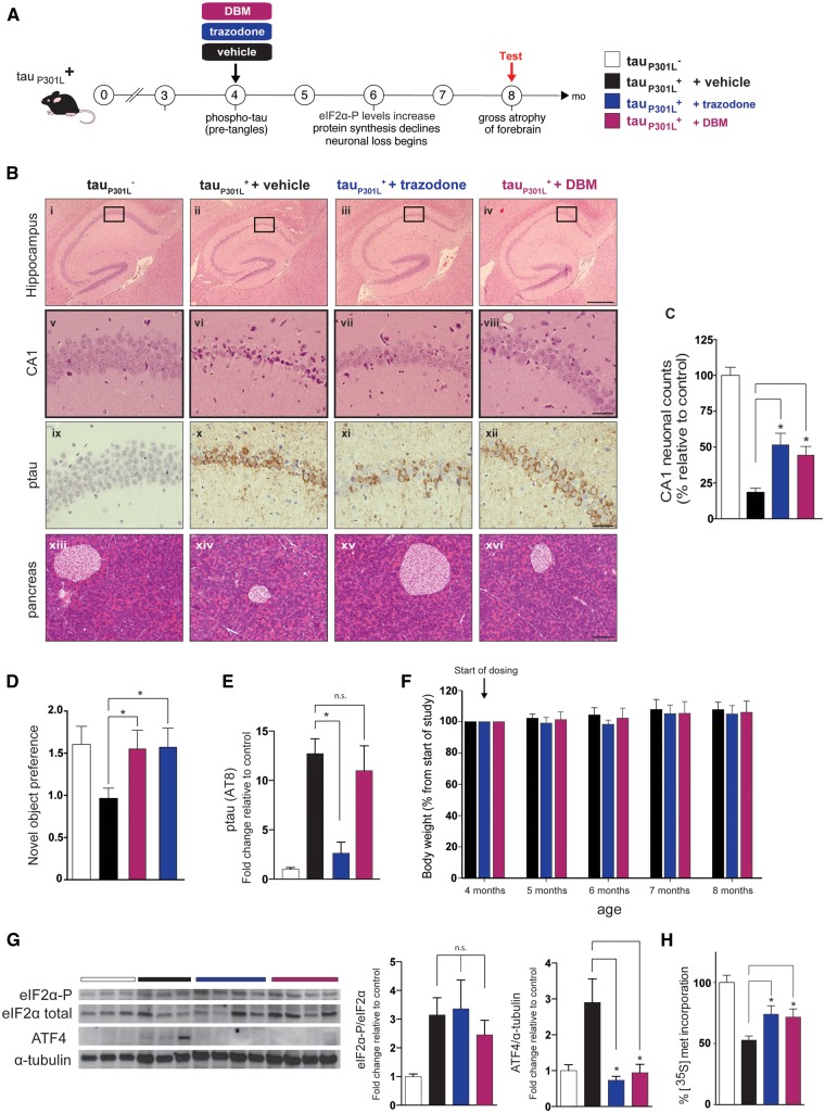
See Mercado and Hetz (doi:10.1093/brain/awx107) for a scientific commentary on this article.Signalling through the PERK/eIF2α-P branch of the unfolded protein response plays a critical role in controlling protein synthesis rates in cells. This pathway is overactivated in brains of patients with Alzheimer's disease and related disorders and has recently emerged as a promising therapeutic target for these currently untreatable conditions. Thus, in mouse models of neurodegenerative disease, prolonged overactivation of PERK/eIF2α-P signalling causes sustained attenuation of protein synthesis, leading to memory impairment and neuronal loss. Re-establishing translation rates by inhibition of eIF2α-P activity, genetically or pharmacologically, restores memory and prevents neurodegeneration and extends survival. However, the experimental compounds used preclinically are unsuitable for use in humans, due to associated toxicity or poor pharmacokinetic properties. To discover compounds that have anti-eIF2α-P activity suitable for clinical use, we performed phenotypic screens on a NINDS small molecule library of 1040 drugs. We identified two compounds, trazodone hydrochloride and dibenzoylmethane, which reversed eIF2α-P-mediated translational attenuation in vitro and in vivo. Both drugs were markedly neuroprotective in two mouse models of neurodegeneration, using clinically relevant doses over a prolonged period of time, without systemic toxicity. Thus, in prion-diseased mice, both trazodone and dibenzoylmethane treatment restored memory deficits, abrogated development of neurological signs, prevented neurodegeneration and significantly prolonged survival. In tauopathy-frontotemporal dementia mice, both drugs were neuroprotective, rescued memory deficits and reduced hippocampal atrophy. Further, trazodone reduced p-tau burden. These compounds therefore represent potential new disease-modifying treatments for dementia. Trazodone in particular, a licensed drug, should now be tested in clinical trials in patients.

Keywords: dementia; drug repurposing; neurodegeneration; therapeutics.


Figures

Figure 1
Figure 1
A screening approach uncovers two partial inhibitors of the UPR. (A) Luciferase expression in CHOP::luciferase cells treated with tunicamycin (Tm) (3 μg/ml) and compounds from primary screen (grey bars) (Supplementary Table 1), ISRIB (turquoise bar), GSK2606414 (green bar) or tunicamycin alone (black bar). ‘Hits’, including DBM (magenta bar) and trazodone (navy bar), repress luciferase expression to similar extent to ISRIB (dotted line). All drugs at 20 μM, except ISRIB, 1 μM; n = 3 and all experiments performed in triplicate. (B) DBM and trazodone inhibit luciferase expression in a dose-dependent manner. Concentrations of tunicamycin and GSK2606414 and n as in A.
Figure 2
Figure 2
Trazodone and DBM inhibit UPR-induced eIF2α-P signalling in vitro. (A) Schematic of the UPR, showing site of action of compounds modulating PERK branch dysregulation. (B) Western blots showing DBM and trazodone reduce ATF4 levels without affecting eIF2α-P levels; bar graphs on right show quantitation. Repeated in triplicate. (C) DBM and trazodone partially restore protein synthesis rates after thapsigargin (1 μM) stress in HEK293 cells, assessed by puromycin incorporation into nascent proteins quantified from western blots. Repeated in triplicate. (D) Trazodone and DBM reduce luciferase expression under control of the ATF4 5’UTR (pATF4), but have no effect when upstream open reading frames in the 5’UTR are removed (pATF4mu), demonstrating an ability to increase ternary complex levels. Levels of pATF4 and pATF4mu normalized to firefly luciferase expressed by the pGL3 plasmid in CHO cells stressed with 1 μM thapsigargin for 6 h. n = 3, repeated in triplicate. (E) XBP1 splicing is unchanged by DBM or trazodone after 3 h tunicamycin stress, determined by RT-PCR, or after 6 h stress and measured by quantitative PCR (F). (G) Full-length ATF6 (flATF6) cleavage to nuclear fragment (nATG6) is not affected by DBM or trazodone. Repeated in triplicate. All bar charts show means ± SEM. *P < 0.05, **P < 0.01, ***P < 0.001, n.s. = non-significant. One-way ANOVA and Tukey’s post hoc test.
Figure 3
Figure 3
Trazodone and DBM are neuroprotective in prion disease. (A) Schematic of prion disease course in tg37+/− mice. (B) Representative images (chosen from n = 10–12) of haematoxylin and eosin stained hippocampal and pancreatic sections, from uninfected controls and prion-infected mice treated with vehicle, trazodone and DBM. Both drugs are markedly neuroprotective (vii, viii compared to vi) and do not harm the pancreas (xi and xii). Scale bars = 400 μm (i–iv), 50 μm (v–viii) 200 μm (ix–xii). (C) Neuronal counts of CA1 region (n = 5 mice for each condition, with three slices counted from each animal). (D) Both drugs prevent loss of object recognition memory and (E) DBM prevents decline in burrowing behaviour at 9 and 10 weeks post-infection (n = 15 for each group) (F) Kaplan-Meier plot shows significantly increased survival with DBM (n = 21) and trazodone (n = 15) compared to vehicle (n = 20) treatment, *P < 0.05, Mantel–Cox analysis used. (G) Weights loss occurs in prion infected mice but not uninfected control, regardless of drug dosing. Normal brain homogenate, treated with trazodone (light blue) or DBM (yellow) n = 5. Prion infected mice treated with vehicle (black, n = 20) trazodone (dark blue, n = 15) or DBM (magenta n = 21). (H) Western blots show high levels of eIF2α-P in prion-disease brains are unaffected by treatment with DBM and trazodone, but ATF4 levels are significantly reduced; bar graphs on right show quantitation relative to loading controls (n = 3; repeated in triplicate). (I) DBM and trazodone restore global protein synthesis rates measured by 35S-met incorporation into hippocampal slices at 10 weeks post-infection (n = 6 per treatment group). (J) Levels of misfolded PrP, PrPSc, detected after PK digestion from brains of terminally sick mice is unaffected by either drug. All bar charts show means ± SEM; *P < 0.05, **P < 0.01, ***P < 0.001, n.s. = non-significant, one-way ANOVA with Tukey’s post hoc test.
Figure 4
Figure 4
Trazodone and DBM are neuroprotective in rTg4510 model of the tauopathy FTD. (A) Schematic of disease progression in rTg4510 tauP301L+ mice. (B) Representative images (chosen from n = 10–12) of hippocampal sections from 8-month-old mice stained with haematoxylin and eosin (i–viii) and pSer202/Thr205-tau (ix–xii) from tauP301L− control mice and tauP301L+ mice treated with vehicle, trazodone or DBM. Scale bars = 400 μm (i–iv), 50 μm (v–xii). (C) Neuronal counts of CA1 region at 8 months (n = 5 for each condition, three consecutive slices). (D) Both drugs prevent loss of object recognition memory (n = 12 for each group). (E) Quantification of p-tau levels measured by the AT100 antibody. Trazodone but not DBM reduces levels. n = 3 mice per group. (F) Trazodone and DBM do not cause weight loss in rTG4510 mice during the 4-month dosing period, n = 10 per group. (G) Protein levels of ATF4 are reduced after treatment with trazodone or DBM compared to vehicle; however, eIF2α-P and eIF2α levels do not change. Repeated in triplicate. (H) DBM and trazodone partially restore global protein synthesis rates measured by 35S-met incorporation into hippocampal slices at 8 months (n = 4 per treatment group). All bar charts show mean ± SEM. *P < 0.05, n.s. = non-significant, using one-way ANOVA and Tukey’s post hoc analysis.

Comment in

Similar articles

Cited by

References

    1. Celardo I, Costa AC, Lehmann S, Jones C, Wood N, Mencacci NEet al. . Mitofusin-mediated ER stress triggers neurodegeneration in pink1/parkin models of Parkinson’s disease. Cell Death Dis 2016; 7: e2271. - PMC - PubMed
    1. Devi L, Ohno M. PERK mediates eIF2 alpha phosphorylation responsible for BACE1 elevation, CREB dysfunction and neurodegeneration in a mouse model of Alzheimer’s disease. Neurobiol Aging 2014; 35: 2272–81. - PMC - PubMed
    1. Dinkova-Kostova AT, Talalay P. Relation of structure of curcumin analogs to their potencies as inducers of Phase 2 detoxification enzymes. Carcinogenesis 1999; 20: 911–14. - PubMed
    1. Frazier MC, Jackson KM, Jankowska-Stephens E, Anderson MG, Harris WB. Proteomic analysis of proteins altered by dibenzoylmethane in human prostatic cancer LNCaP cells. Proteomics 2004; 4: 2814–21. - PubMed
    1. Halliday M, Mallucci GR. Modulating the unfolded protein response to prevent neurodegeneration and enhance memory. Neuropathol Appl Neurobiol 2015; 41: 414–27. - PMC - PubMed

MeSH terms