. 2017 May 1;96(5):993-1006.
doi: 10.1093/biolre/iox029.

IFT25, an intraflagellar transporter protein dispensable for ciliogenesis in somatic cells, is essential for sperm flagella formation

Affiliations

IFT25, an intraflagellar transporter protein dispensable for ciliogenesis in somatic cells, is essential for sperm flagella formation

Hong Liu et al. Biol Reprod. .

Abstract

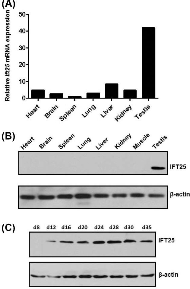
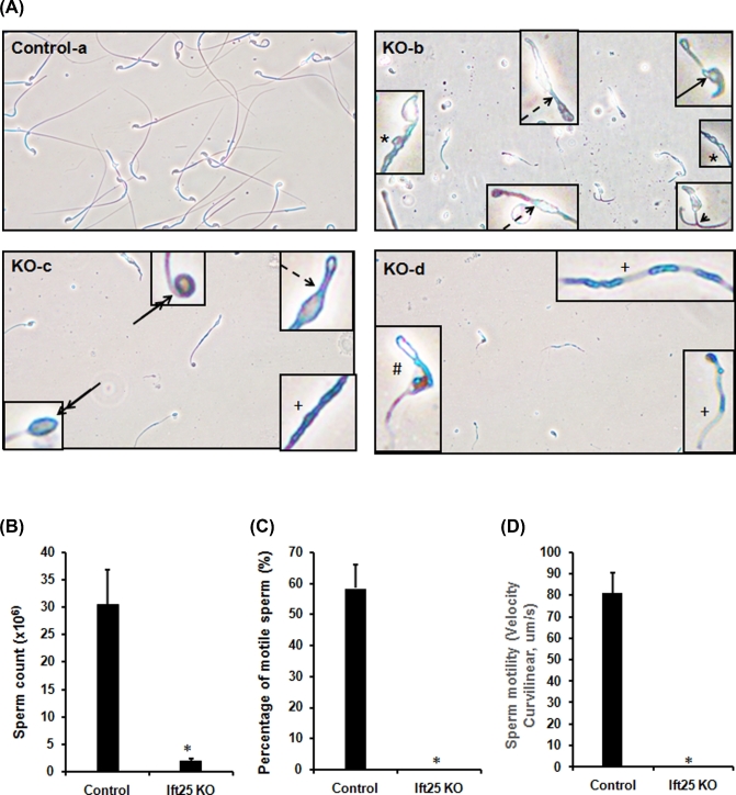
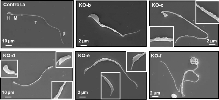
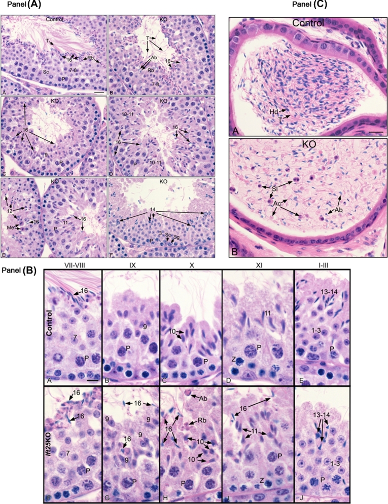
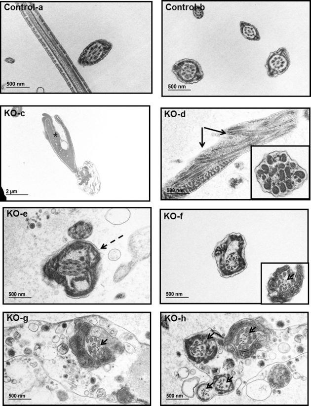
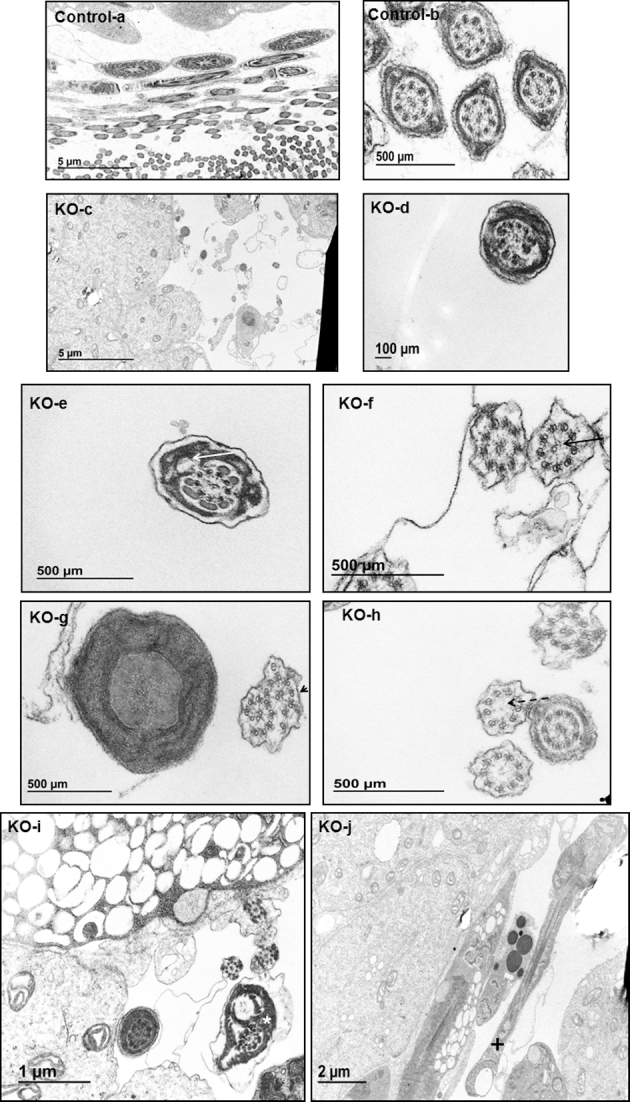
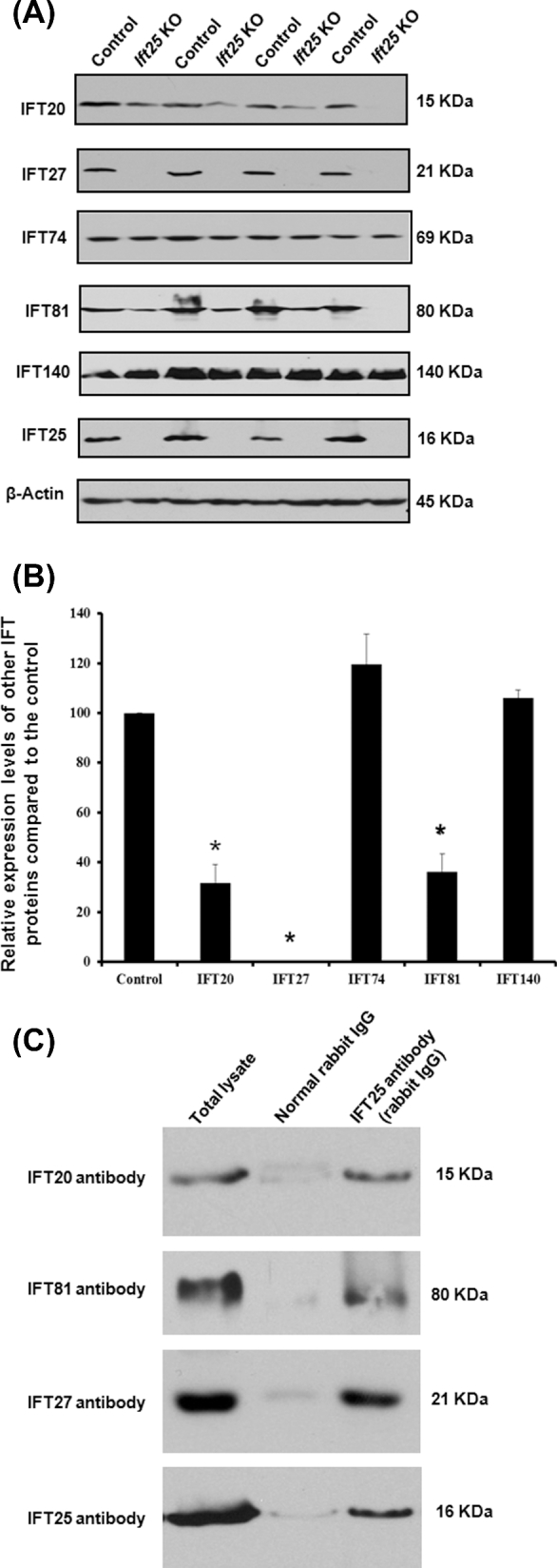
Intraflagellar transport (IFT) is a conserved mechanism essential for the assembly and maintenance of most eukaryotic cilia and flagella. However, IFT25, a component of the IFT complex, is not required for the formation of cilia in somatic tissues. In mice, the gene is highly expressed in the testis, and its expression is upregulated during the final phase when sperm flagella are formed. To investigate the role of IFT25 in sperm flagella formation, the gene was specifically disrupted in male germ cells. All homozygous knockout mice survived to adulthood and did not show any gross abnormalities. However, all homozygous knockout males were completely infertile. Sperm numbers were reduced and these sperm were completely immotile. Multiple morphological abnormalities were observed in sperm, including round heads, short and bent tails, with some tails showing branched flagella and others with frequent abnormal thicknesses, as well as swollen tips of the tail. Transmission electron microscopy revealed that flagellar accessory structures, including the fibrous sheath and outer dense fibers, were disorganized, and most sperm had also lost the "9+2" microtubule structure. In the testis, IFT25 forms a complex with other IFT proteins. In Ift25 knockout testes, IFT27, an IFT25 binding partner, was missing, and IFT20 and IFT81 levels were also reduced. Our findings suggest that IFT25, although not necessary for the formation of cilia in somatic cells, is indispensable for sperm flagellum formation and male fertility in mice.

Keywords: ciliogenesis; flagellogenesis; germ cells; intraflagellar transport; spermiogenesis.


Figures

Figure 1.
Figure 1.
IFT25 is highly expressed in the testis and the protein is upregulated during spermiogenesis. (A) Analysis of relative Ift25 mRNA expression in adult mouse tissues by qPCR. Ift25 mRNA expression levels were normalized by Gapdh. (B) IFT25 protein expression in adult mouse tissues. Notice that IFT25 was detected only in the testis when less-sensitive Pico system was used. (C) Testicular IFT25 expression during the first wave of spermatogenesis. Notice that the protein was expressed from day 12, when germ cells enter meiosis phase. It appears to be upregulated from day 24, when germ cells enter the final phase, spermiogenesis.
Figure 2.
Figure 2.
Disruption of Ift25 gene in germ cells leads to abnormal sperm morphology associated with significantly reduced sperm count and sperm immobility. (A) Examination of epididymal sperm from 3–4-month-old control (a) and conditional Ift25 KO mice (b–d) by light microscopy. Notice that under the same dilution, compared to the control, much cell debris was present, and very few sperm were seen in the conditional Ift25 KO mice. Sperm from the KO mice usually had shorter (black arrow in b) or branched tails (black arrowhead in b). Some sperm had longer tails than the others, but vacuoles were present along the tails (dashed arrows in b and c). Double-arrow lines point to the sperm with swollen tails; the asterisk showed the sperm flagella with different thickness; the plus indicated the sperm flagella with different light density, probably stuck IFT particles inside the developing tails; the hash indicates a spermatozoon with a bent tail. (B) Sperm count is significantly reduced in the KO mice. (C and D). Sperm motile in 3–4-month-old control and the KO mice analyzed in PBS. Notice that no sperm showed motility in the KO mice.
Figure 3.
Figure 3.
Examination of epididymal sperm by SEM. (a) Representative image of epididymis sperm from a 4-month-old control mouse with normal morphology. The sperm has normally developed midpiece (M), a long and smooth tail (T), and nicely condensed head (H). (b–f) Representative images of epididymal sperm from a 4-month-old KO mouse. Variety of abnormal morphology of sperm from the KO mice was present. Some have short and kinked tails (b), some have round (c), and other misshaped heads (upper right inserts in d and e); some have relatively longer but uneven tails (the two inserts in c, low right insert in d); some have abnormal tail tips (left insert in d, and low insert in e). Some sperm have branched tails (f).
Figure 4.
Figure 4.
Testicular and epididymal histology of adult control and Ift25 KO mice. Panel A: (A) control testis showing stage VII-VIII. Step 16 spermatids line the lumen, with their long tails (T) extending parallel into the lumen. Residual bodies (Rb) of left over cytoplasm are forming between the sperm heads and the step 7–8 round spermatids. Sc, Sertoli cell; P, pachytene spermatocytes. Bar = 20 μm. (B–F) Ift25 KO mice testes. (B) Stage VII-VIII, showing abnormal tail (T) formation from the step 16 spermatids. Abnormal step 16 spermatid head (Ab) are present above the nuclei of step 7–8 round spermatids. (C) Stage IX, showing failure of spermiation with step 16 spermatid head being retained within the seminiferous epithelium and among the step 9 spermatids that are just beginning to change shape. P, pachytene spermatocytes. (D) Stage X-XI, showing step 16 spermatid head being retained in the epithelium along with step 10–11 spermatids having large variation in shape, with the beginning of abnormal formations. P, pachytene spermatocytes. (E) Stage XII on the left and stage XI on the right. Step 16 spermatid head are seen retained in the epithelium and abnormal step 11–12 spermatids are seen due to alterations in nuclear shape. Me, necrotic spermatocytes in meiotic division. (F) Stage III, showing numerous step 14 elongating spermatid heads. Step 3 round spermatid nuclei are normal in appearance. Sc, Sertoli cell; P, pachytene spermatocyte; Sg, spermatogonium. Panel B. Control (A–E) and Ift25 KO (F–J). Round spermatids are indicated by 1–7. Elongating spermatids are indicated by 9–16. P, pachytene spermatocyte; Z, zygotene spermatocyte. Bar = 10 μm for all photos. Stage VII-VIII: control and Ift25 KO step 7 round spermatids are normal. Elongated spermatids step 16 in control have thin, highly condensed nuclei that are aligned at the lumen for spermiation, but in the KO step 16 spermatids show abnormal alignment and head formations. P, pachytene spermatocytes. Bar = 10 μm for all photos. Stage IX: step 9 spermatids in control show the beginning of head elongation. In the Ift25 KO some step 9 cells are normal, but there is an inconsistent change in spermatid head elongation. There are also step 16 spermatid heads that were not released, an indication of failure of spermiation. Stage X: step 10 spermatids in controls have elongated and show the typical protrusion of the condensing head. However, step 10 spermatid heads in Ift25 KO males show significant abnormality in shape and appear disorganized in the epithelium. Numerous step 16 spermatids remain attached to the epithelium after failure of spermiation in stage VIII. An abnormal aggregate of step 16 spermatid heads (Ab) is seen near the lumen. Residual bodies (Rb) are also retained abnormally near the lumen. Stage XI: control step 11 elongating spermatids are well formed in the seminiferous epithelium above the large pachytene spermatocytes (P). In Ift25 KO, step 11 cell nuclei show abnormal shapes, and the heads of step 16 spermatids remain attached, rather than being released into the lumen. Stage I-III: round spermatids (1-3) appear normal in shape and numbers in both control and Ift25 KO testes. In control, the elongating spermatids steps 13–14 have condensed chromatin heads that are positioned throughout the epithelium. In Ift25 KO, step 13–14 cells show multiple head abnormal shapes, which appeared first in earlier stages. Panel C: cauda epididymis from control and Ift25 KO mice. Control sperm are highly concentrated in the cauda region, showing the head (Hd) and tails (T). In Ift25 KO, the cauda epididymis lumen is filled with abnormal sperm heads (Ab), abnormal cytoplasm attached to sperm tails (Ac), sloughed spermatids (Sl), numerous detached sperm heads and abnormal tails.
Figure 5.
Figure 5.
Analysis of epididymal sperm of the control and Ift25 KO mice by TEM. TEM images of epididymal sperm from 4-month-old control and KO mice. Sperm from the control mice show normal ultrastructure (a, b). However, most sperm from the Ift25 KO demonstrate multiple abnormalities, including abnormal head (asterisk in c), disorganized microtubule array (arrows in d), distorted membranes (dashed arrow in e); some sperm lose “9+2” core axoneme structure (arrow heads in f, g, h); some have disorganized (insert in d), or missing accessary structures, including ODF and fibrous sheath (g, h).
Figure 6.
Figure 6.
Analysis of testicular sperm of the control and Ift25 KO mice by TEM. TEM images of testicular sperm from 4-month-old control and KO mice. In the control mice, sperm were easily discovered in the lumen of seminiferous tubules (a), and the ultrastructure was normal (b). However, few sperm were discovered in the KO (c), and the sperm usually have disorganized axoneme structure (d). The white arrow in “e” points to an axoneme missing several ODFs; the black arrow in “f” points to an axoneme missing one of the two central microtubule; the arrow head in “g” points to an axoneme with extra microtubules; the dashed arrow in “h” points to an axoneme missing the entire central microtubules; the white asterisk in “i” shows a sperm with distorted membrane; and the plus in “j” shows a sperm with a vacuole in the tail.
Figure 7.
Figure 7.
Regulation of other IFT proteins in absence of IFT25 in male germ cells. (A) Representative western blot results using the indicated IFT antibodies. Notice that IFT27, the IFT25 binding partner, is missing in the testes of the Ift25 KO mice. Testicular expression of IFT20 and IFT81 were also reduced in the KO. (B) Statistical analysis of relative expression of the IFT proteins normalized by β-actin. There is no difference in expression levels of IFT74 and IFT140 between the controls and the Ift25 KO mice. IFT20 and IFT81 expression levels are significantly reduced in the KO. (C) Both IFT20 and IFT81 are present in the IFT25 complex in mouse testis. Testicular extract was pulled down with anti-IFT25 antibody, and western blottings were conducted using anti-IFT25, anti-IFT20, and anti-IFT81 antibodies.

Similar articles

Cited by

References

    1. Malicki JJ, Johnson CA. The Cilium: cellular antenna and central processing unit. Trends Cell Biol 2017; 27:126–140. - PMC - PubMed
    1. Rohatgi R, Snell WJ. The ciliary membrane. Curr Opin Cell Biol 2010; 22:541–546. - PMC - PubMed
    1. Emmer BT, Maric D, Engman DM. Molecular mechanisms of protein and lipid targeting to ciliary membranes. J Cell Sci 2010; 123:529–536. - PMC - PubMed
    1. Pedersen LB, Mogensen JB, Christensen ST. Endocytic control of cellular signaling at the primary cilium. Trends Biochem Sci 2016; 41:784–797. - PubMed
    1. Prasad RM, Jin X, Nauli SM. Sensing a sensor: identifying the mechanosensory function of primary cilia. Biosensors (Basel) 2014; 4:47–62. - PMC - PubMed

MeSH terms

Substances