Review
. 2017 Jul 1;27(7):601-618.
doi: 10.1093/glycob/cwx034.

Biological functions of fucose in mammals

Affiliations
Review

Biological functions of fucose in mammals

Michael Schneider et al. Glycobiology. .

Abstract

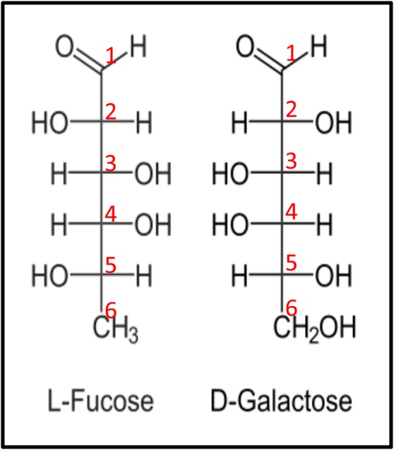
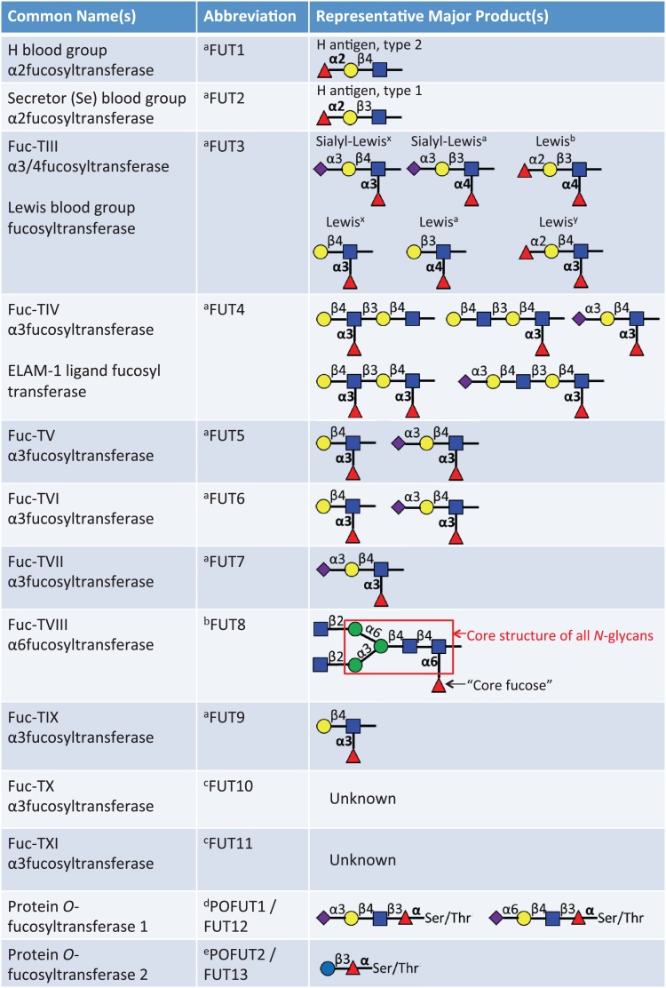
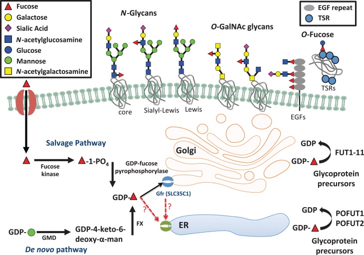
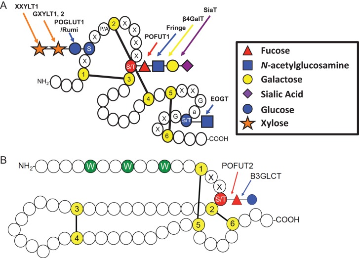
Fucose is a 6-deoxy hexose in the l-configuration found in a large variety of different organisms. In mammals, fucose is incorporated into N-glycans, O-glycans and glycolipids by 13 fucosyltransferases, all of which utilize the nucleotide-charged form, GDP-fucose, to modify targets. Three of the fucosyltransferases, FUT8, FUT12/POFUT1 and FUT13/POFUT2, are essential for proper development in mice. Fucose modifications have also been implicated in many other biological functions including immunity and cancer. Congenital mutations of a Golgi apparatus localized GDP-fucose transporter causes leukocyte adhesion deficiency type II, which results in severe developmental and immune deficiencies, highlighting the important role fucose plays in these processes. Additionally, changes in levels of fucosylated proteins have proven as useful tools for determining cancer diagnosis and prognosis. Chemically modified fucose analogs can be used to alter many of these fucose dependent processes or as tools to better understand them. In this review, we summarize the known roles of fucose in mammalian physiology and pathophysiology. Additionally, we discuss recent therapeutic advances for cancer and other diseases that are a direct result of our improved understanding of the role that fucose plays in these systems.

Keywords: cancer; development; fucose; fucosyltransferase; immunology.


Figures

Fig. 1.
Fig. 1.
Fischer projection formula of l-fucose. The six carbons of fucose are numbered. Note that most naturally occurring sugars, such as galactose, are present in the d-configuration, as can be determined by the arrangement of the hydroxyl group bound to the C-5 carbon. Note further that the C-6 carbon of l-fucose lacks a hydroxyl group present at the C-6 position of d-galactose. l-Fucose can also be described as 6-deoxy-l-galactose. This figure is available in black and white in print and in color at Glycobiology online.
Fig. 2.
Fig. 2.
List of 13 known fucosyltransferases in humans. Major representative products of each fucosyltransferase are listed. The linkage of the fucose added by each enzyme appears in bold.aThese enzymes can add fucose to oligosaccharide chains on glycolipids, N-glycans or mucin O-glycans. bThis enzyme only adds the core fucose to N-glycans. cThese are putative α3-fucosyltransferases. Acceptor substrates have not been clearly defined. dThis modification is only observed in O-fucose consensus sequences on EGF repeats (C2XXXX(S/T)C3), see Figure 4A. eThis modification is only observed in O-fucose consensus sequences on TSRs (C1−2XX(S/T)C2−3), see Figure 4B. This figure is available in black and white in print and in color at Glycobiology online.
Fig. 3.
Fig. 3.
Fucose metabolism pathways and variation in types of fucosylated glycans. This figure illustrates the de novo fucose synthesis pathway, which converts GDP-mannose to GDP-fucose and the fucose salvage pathway, which converts free fucose taken up from outside the cell to GDP-fucose. GDP-fucose can then be taken up into the Golgi apparatus by the GDP-fucose transporter (SLC35C1) and possibly into the ER by an as yet unknown transporter. Proteins are then modified with GDP-fucose and other carbohydrates within the Golgi and ER and can then be secreted or expressed on the cell surface. This figure is available in black and white in print and in color at Glycobiology online.
Fig. 4.
Fig. 4.
Key features of EGF repeats and TSRs. (A) Cartoon showing a single EGF repeat. Each circle represents one amino acid. Conserved cysteines (yellow) are numbered and disulfide bonds are indicated. O-Glucose and O-GlcNAc sites are shaded blue and the O-fucose site is shaded red. Enzymes responsible for the addition of each sugar are indicated. Modified from Rana and Haltiwanger (2011). Used with permission. Elsevier. (B) Cartoon showing a typical TSR. Conserved cysteines (yellow) and disulfide bonds are indicated. C-Mannose sites are shown in green and the O-fucose site is shaded red. (S) Serine; (T) Threonine; (G) Glycine; (W) Tryptophan; (X) any amino acid, (a) any aromatic amino acid. Modified with permission from Haltiwanger (2004). ©Elsevier. This figure is available in black and white in print and in color at Glycobiology online.

Similar articles

Cited by

References

    1. Abd El-Aziz MM, Barragan I, O'Driscoll CA, Goodstadt L, Prigmore E, Borrego S, Mena M, Pieras JI, El-Ashry MF, Safieh LA et al. . 2008. EYS, encoding an ortholog of Drosophila spacemaker, is mutated in autosomal recessive retinitis pigmentosa. Nat Genet. 40:1285–1287. - PMC - PubMed
    1. Adamczyk B, Tharmalingam T, Rudd PM. 2012. Glycans as cancer biomarkers. Biochim Biophys Acta (BBA)-Gen Subjects. 1820:1347–1353. - PubMed
    1. Ahram D, Sato TS, Kohilan A, Tayeh M, Chen S, Leal S, Al-Salem M, El-Shanti H. 2009. A homozygous mutation in ADAMTSL4 causes autosomal-recessive isolated ectopia lentis. Am J Human Genetics. 84:274–278. - PMC - PubMed
    1. Al-Shareffi E, Chaubard JL, Leonhard-Melief C, Wang SK, Wong CH, Haltiwanger RS. 2013. 6-Alkynyl fucose is a bioorthogonal analog for O-fucosylation of epidermal growth factor-like repeats and thrombospondin type-1 repeats by protein O-fucosyltransferases 1 and 2. Glycobiology. 23:188–198. - PMC - PubMed
    1. Aldahmesh MA, Alshammari MJ, Khan AO, Mohamed JY, Alhabib FA, Alkuraya FS. 2013. The syndrome of microcornea, myopic chorioretinal atrophy, and telecanthus (MMCAT) is caused by mutations in ADAMTS18. Hum Mutat. 34:1195–1199. - PubMed

Publication types