. 2017 Jul 1;26(13):2472-2479.
doi: 10.1093/hmg/ddx142.

Evidence of nuclei-encoded spliceosome mediating splicing of mitochondrial RNA

Affiliations

Evidence of nuclei-encoded spliceosome mediating splicing of mitochondrial RNA

Roberto H Herai et al. Hum Mol Genet. .

Erratum in

Abstract

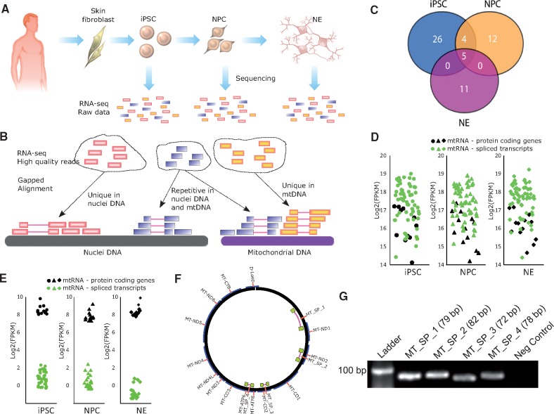
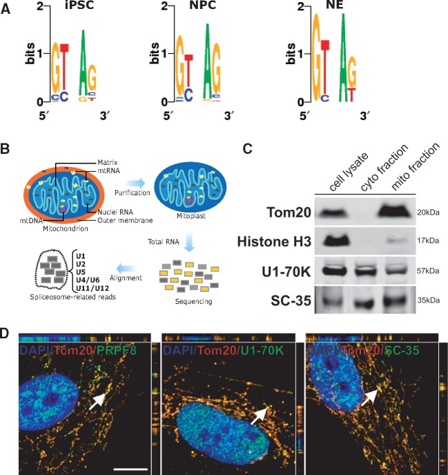
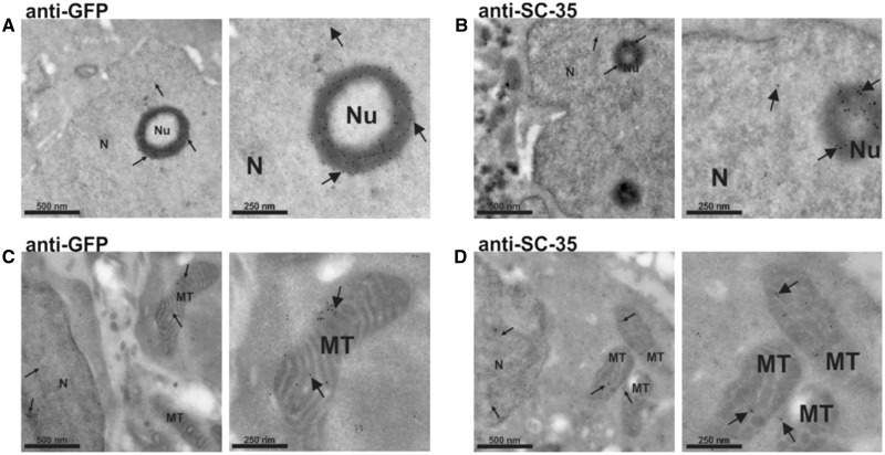
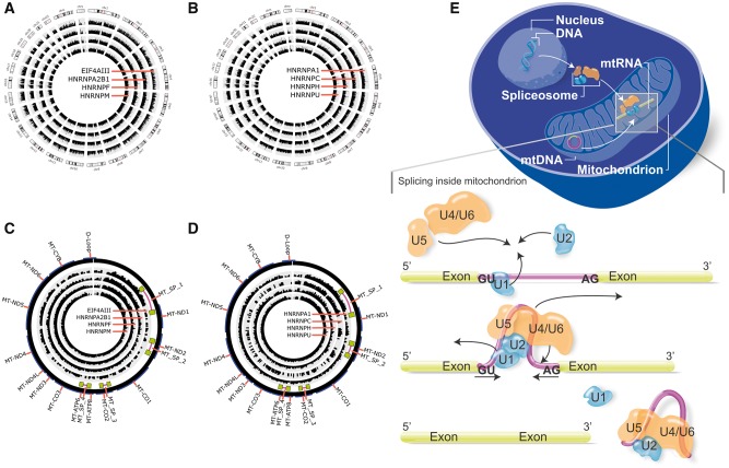
Mitochondria are thought to have originated as free-living prokaryotes. Mitochondria organelles have small circular genomes with substantial structural and genetic similarity to bacteria. Contrary to the prevailing concept of intronless mitochondria, here we present evidence that mitochondrial RNA transcripts (mtRNA) are not limited to policystronic molecules, but also processed as nuclei-like transcripts that are differentially spliced and expressed in a cell-type specific manner. The presence of canonical splice sites in the mtRNA introns and of core components of the nuclei-encoded spliceosome machinery within the mitochondrial organelle suggest that nuclei-encoded spliceosome can mediate splicing of mtRNA.


Figures

Figure 1
Figure 1
Analysis of spliced mitochondrial RNA transcripts. (A) Reprogramming of HPSF into human induced pluripotent stem cells (iPSC). The iPSC were then differentiated into neuronal progenitor cells (NPC) and postmitotic neurons (NE). RNA-seq data from these three cell types were analyzed. (B) High quality reads from RNA-seq were aligned against the human mitochondrial DNA and to the human genomic DNA to identify non-redundant mtRNA spliced reads. (C) Venn diagram showing the number of common or exclusive mitochondrial spliced transcripts in iPSC, NPC and NE. (D) Expression levels (including repetitive reads) in FPKM of the 13 known protein-coding genes from mitochondria and the respective spliced transcripts found in iPSC, NPC and NE. (E) Expression levels (excluding repetitive and non-spliced reads) in FPKM of the 13 known protein-coding genes from mitochondria and the respective spliced transcripts found in iPSC, NPC and NE. (F) Mitochondrial DNA locus of the four spliced transcripts identified and tested by PCR. (G) PCR amplification products of the four spliced mtRNA transcripts observed in HPSF.
Figure 2
Figure 2
Presence of nuclei-encoded spliceosome proteins within mitochondria. (A) Analysis of the dinucleotide motifs on the 5′ and 3′ ends of introns in spliced mtRNA from iPSC, NPC and NE. (B) Schematic of the detection of nucleus-encoded spliceosome in mitoplast RNA-seq data. (C) Western blot analysis for Tom20, Histone H3, U1-70K and SC-35 in total cell lysate compared with equal protein amounts of cytosolic- and mithochondrial-enriched fractions from HPSF. (D) Co-localization of spliceosomal proteins and mitochondria in HPSF. Cells were fixed and stained for the mitochondrial translocase Tom20, and three spliceosomal proteins PRPF8, U1-70k and SC-35. White arrows indicate random sites chosen for three-dimensional-z-stack analysis. Scale bar, 10 μm (images correspond to merged panels showed on Supplementary Material Figure S1C).
Figure 3
Figure 3
Electron microscopic distribution of SC-35 in HEK293 cells transfected with a SC35-GFP-flag. Cells were stained with a primary antibody against GFP (A and C) or SC-35 (B and D) protein, and a secondary antibody conjugated to 10 nm colloidal gold. Proteins are diffusely distributed throughout the nucleoplasm. A cluster of splicing factors is also concentrated in a ring shape structure around the nucleolus (A and B). Regions of SC-35 immunostaining extend out the nucleus and the signal is clearly found within the mitochondria (C and D), suggesting that the protein is being imported into this organelle. Images on the right panels are higher magnifications from those on the left panels. Arrows highlight the location of some gold particles. N: nucleus; Nu: nucleolus; MT: mitochondria. Scale bars are indicated.
Figure 4
Figure 4
Representation of core spliceosome proteins in human somatic and non-somatic chromosomes, and an overview of the mtRNA splicing mediated nuclei-encoded spliceosome proteins. (A and C) Clip-seq data of the core spliceosome proteins ELF4AIII, HNRNPA2B1, HNRNPF, HNRNPM occupying mitochondrial RNA transcripts and nuclei RNA transcripts, respectively; (B and D) Clip-seq data of the core spliceosome proteins HNRNPA1, HNRNPC, HNRNPH, HNRNPU occupying mitochondrial RNA transcripts and nuclei RNA transcripts, respectively. Data coverage (internal histograms) were normalized according to sample size and a log base 2 scale was applied over read coverage, ranging from 0 to 100 for both mitochondrial DNA (circular genome) and nuclei DNA (linear chromosomes 1-22, X and Y). (E) Schematic model of nuclei-encoded spliceosome moving from nuclei to cytosol, and then imported into mitochondrial compartments to occupy and splice mitochondrial RNA transcripts.

Similar articles

Cited by

References

    1. Gray M.W., Burger G., Lang B.F. (2001) The origin and early evolution of mitochondria. Genome Biol., 2, REVIEWS1018.. - PMC - PubMed
    1. Asin-Cayuela J., Gustafsson C.M. (2007) Mitochondrial transcription and its regulation in mammalian cells. Trends Biochem. Sci., 32, 111–117. - PubMed
    1. Timmis J.N., Ayliffe M.A., Huang C.Y., Martin W. (2004) Endosymbiotic gene transfer: organelle genomes forge eukaryotic chromosomes. Nat. Rev. Genet., 5, 123–135. - PubMed
    1. Gray M.W. (1999) Mitochondrial Evolution. Science, 283, 1476–1481. - PubMed
    1. Mercer T.R., Neph S., Dinger M.E., Crawford J., Smith M.A., Shearwood A.-M.J., Haugen E., Bracken C.P., Rackham O., Stamatoyannopoulos J.A.. et al. (2011) The human mitochondrial transcriptome. Cell, 146, 645–658. - PMC - PubMed