

Protective Effect of Mesenchymal Stem Cells Against the Development of Intracranial Aneurysm Rupture in Mice
- PMID: 28431181
- PMCID: PMC6257015
- DOI: 10.1093/neuros/nyx172
Protective Effect of Mesenchymal Stem Cells Against the Development of Intracranial Aneurysm Rupture in Mice
Abstract
Background: Mesenchymal stem cells (MSCs) are multipotent stem or stromal cells found in multiple tissues. Intravenous MSC injections have been used to treat various diseases with an inflammatory component in animals and humans. Inflammation is emerging as a key component of pathophysiology of intracranial aneurysms. Modulation of inflammation by MSCs may affect sustained inflammatory processes that lead to aneurysmal rupture.
Objective: To assess the effect of MSCs on the development of aneurysm rupture using a mouse model.
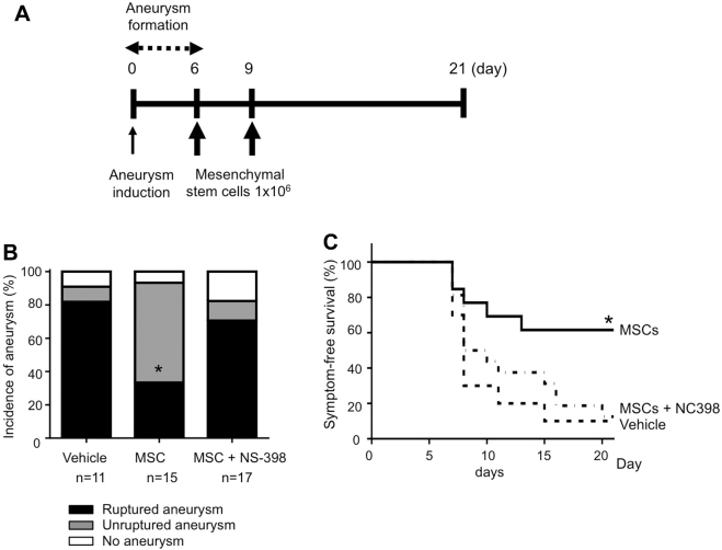
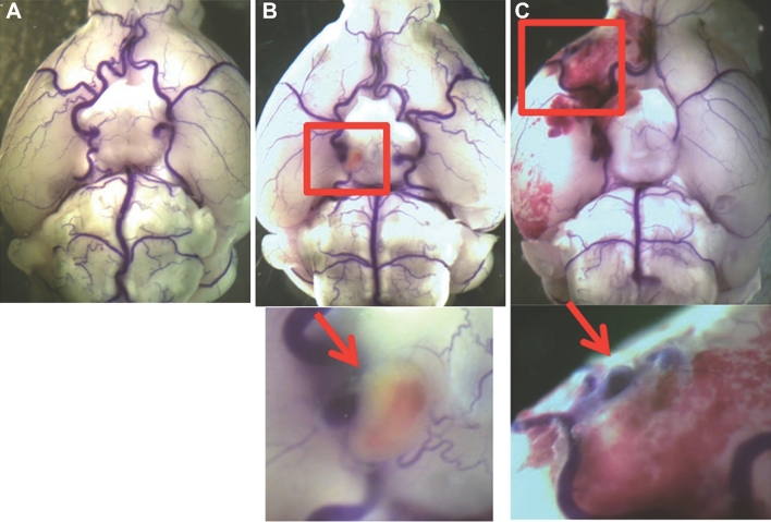
Methods: Intracranial aneurysms were induced with a combination of a single elastase injection into the cerebrospinal fluid and deoxycorticosterone acetate salt-induced hypertension in mice. We administered allogeneic bone marrow-derived MSCs or vehicle, 6 and 9 d after aneurysm induction.
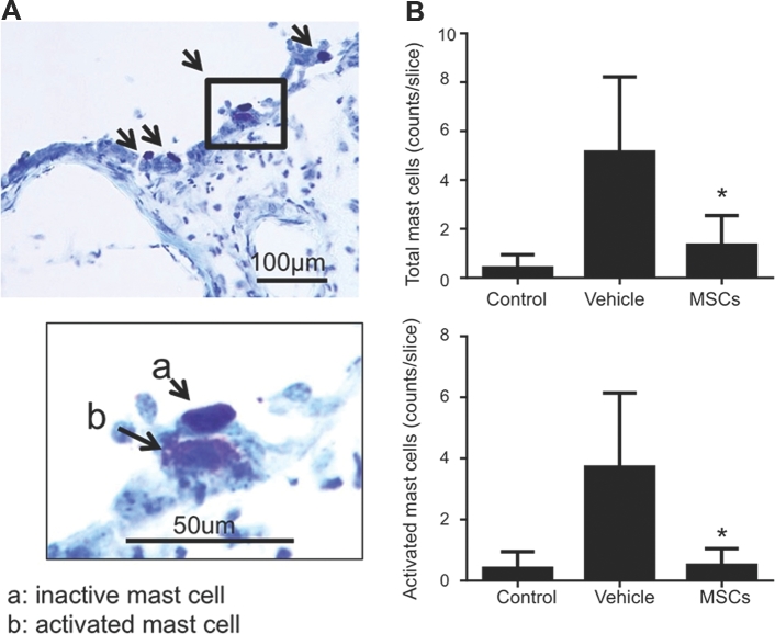
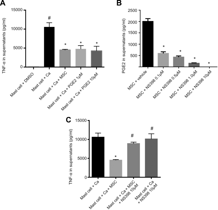
Results: MSC administration significantly reduced rupture rate (vehicle control vs MSCs, 90% vs 36%; P < .05). In cell culture experiments with an MSC and mast cell coculture, MSCs stabilized mast cells through cyclooxygenase-2 (COX-2)-dependent production of prostaglandin E2, thereby reducing the release of proinflammatory cytokines from mast cells. Pretreatment of MSCs with COX-2 inhibitor, NS-398, abolished the protective effect of MSCs against the development of aneurysm rupture.
Conclusion: Intravenous administration of MSCs after aneurysm formation prevented aneurysmal rupture in mice. The protective effect of MSCs against the development of aneurysm rupture appears to be mediated in part by the stabilization of mast cells by MSCs.
Keywords: Intracranial aneurysm; Mesenchymal stem cells; Stroke; Subarachnoid hemorrhage.
Copyright © 2017 by the Congress of Neurological Surgeons
Figures
Similar articles
-
Human Mesenchymal Stem Cell-Derived Microvesicles Prevent the Rupture of Intracranial Aneurysm in Part by Suppression of Mast Cell Activation via a PGE2-Dependent Mechanism.Stem Cells. 2016 Dec;34(12):2943-2955. doi: 10.1002/stem.2448. Epub 2016 Jul 8. Stem Cells. 2016. PMID: 27350036 Free PMC article.
-
Mast Cell Promotes the Development of Intracranial Aneurysm Rupture.Stroke. 2020 Nov;51(11):3332-3339. doi: 10.1161/STROKEAHA.120.030834. Epub 2020 Oct 6. Stroke. 2020. PMID: 33019897 Free PMC article.
-
Prevention Effect of Antiplatelets on Aneurysm Rupture in a Mouse Intracranial Aneurysm Model.Cerebrovasc Dis. 2018;45(3-4):180-186. doi: 10.1159/000487812. Epub 2018 Apr 3. Cerebrovasc Dis. 2018. PMID: 29614486
-
Potential role of aspirin in the prevention of aneurysmal subarachnoid hemorrhage.Cerebrovasc Dis. 2015;39(5-6):332-42. doi: 10.1159/000381137. Epub 2015 May 7. Cerebrovasc Dis. 2015. PMID: 25967073 Free PMC article. Review.
-
Pharmaceutical Modulation of Intracranial Aneurysm Development and Rupture.J Clin Med. 2024 Jun 5;13(11):3324. doi: 10.3390/jcm13113324. J Clin Med. 2024. PMID: 38893035 Free PMC article. Review.
Cited by
-
Exosomes in Vascular/Neurological Disorders and the Road Ahead.Cells. 2024 Apr 12;13(8):670. doi: 10.3390/cells13080670. Cells. 2024. PMID: 38667285 Free PMC article. Review.
-
Mast Cells Mediate Inflammatory Injury and Aggravate Neurological Impairment in Experimental Subarachnoid Hemorrhage Through Microglial PAR-2 Pathway.Front Cell Neurosci. 2021 Sep 27;15:710481. doi: 10.3389/fncel.2021.710481. eCollection 2021. Front Cell Neurosci. 2021. PMID: 34646122 Free PMC article.
-
The Role of Mast Cells in Stroke.Cells. 2019 May 10;8(5):437. doi: 10.3390/cells8050437. Cells. 2019. PMID: 31083342 Free PMC article. Review.
-
Bioactive refinement for endosaccular treatment of intracranial aneurysms.Neuroradiol J. 2021 Dec;34(6):534-541. doi: 10.1177/19714009211024631. Epub 2021 Jul 1. Neuroradiol J. 2021. PMID: 34210195 Free PMC article. Review.
-
Pharmacological inhibition of STAT3 by BP-1-102 inhibits intracranial aneurysm formation and rupture in mice through modulating inflammatory response.Pharmacol Res Perspect. 2021 Feb;9(1):e00704. doi: 10.1002/prp2.704. Pharmacol Res Perspect. 2021. PMID: 33474811 Free PMC article.
References
-
- Connolly ES Jr, Rabinstein AA, Carhuapoma JR et al. . Guidelines for the management of aneurysmal subarachnoid hemorrhage: a guideline for healthcare professionals from the American Heart Association/american Stroke Association. Stroke. 2012;43(6):1711-1737. - PubMed
MeSH terms
Grants and funding
LinkOut - more resources
Full Text Sources
Other Literature Sources
Medical
Research Materials
Miscellaneous
