. 2017 Apr 20;169(3):497-509.e13.
doi: 10.1016/j.cell.2017.03.044.

Spontaneous Chitin Accumulation in Airways and Age-Related Fibrotic Lung Disease

Affiliations

Spontaneous Chitin Accumulation in Airways and Age-Related Fibrotic Lung Disease

Steven J Van Dyken et al. Cell. .

Abstract

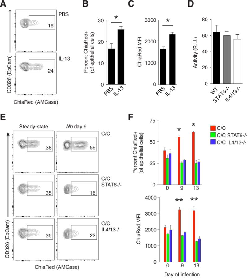
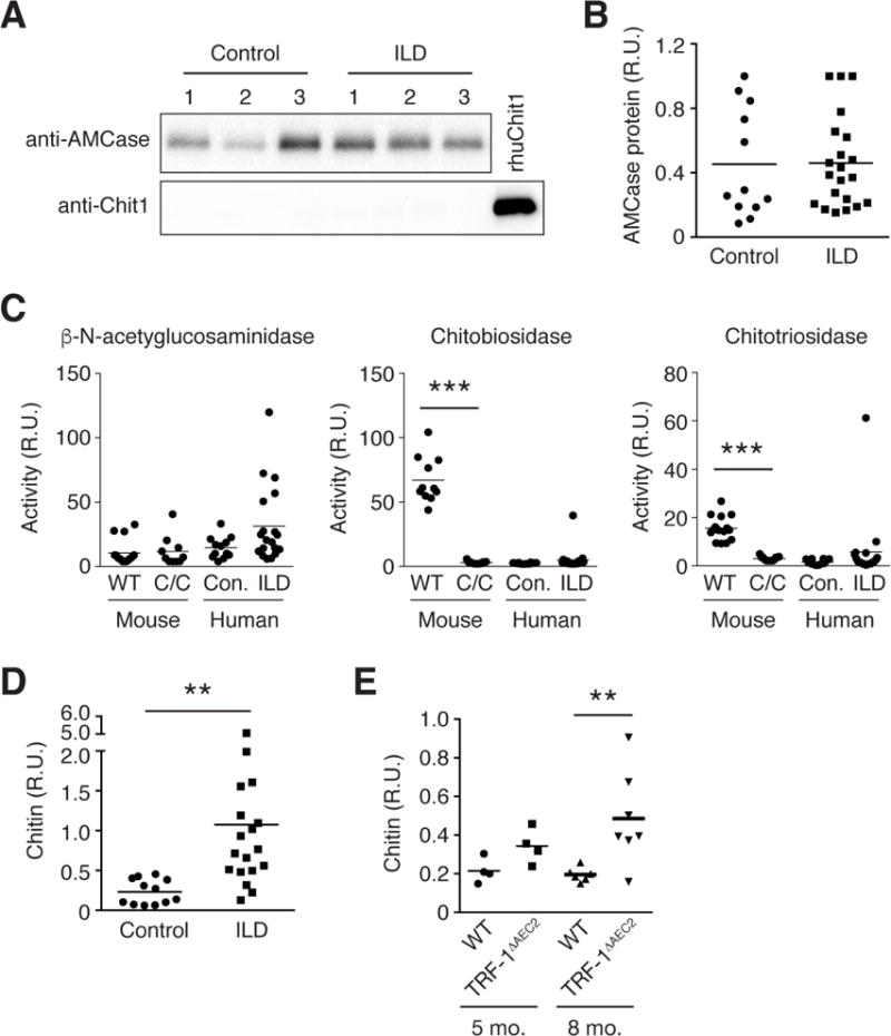
The environmentally widespread polysaccharide chitin is degraded and recycled by ubiquitous bacterial and fungal chitinases. Although vertebrates express active chitinases from evolutionarily conserved loci, their role in mammalian physiology is unclear. We show that distinct lung epithelial cells secrete acidic mammalian chitinase (AMCase), which is required for airway chitinase activity. AMCase-deficient mice exhibit premature morbidity and mortality, concomitant with accumulation of environmentally derived chitin polymers in the airways and expression of pro-fibrotic cytokines. Over time, these mice develop spontaneous pulmonary fibrosis, which is ameliorated by restoration of lung chitinase activity by genetic or therapeutic approaches. AMCase-deficient epithelial cells express fibrosis-associated gene sets linked with cell stress pathways. Mice with lung fibrosis due to telomere dysfunction and humans with interstitial lung disease also accumulate excess chitin polymers in their airways. These data suggest that altered chitin clearance could exacerbate fibrogenic pathways in the setting of lung diseases characterized by epithelial cell dysfunction.

Keywords: AMCase; age-related disease; chitin; chitinase; epithelium; interleukins; interstitial lung disease; polysaccharide; pulmonary fibrosis.


Figures

FIGURE 1
FIGURE 1. Airway epithelial cells produce active AMCase
(A) AMCase (Chia1) gene targeting strategy and map of ChiaRed allele. (B) Flow cytometry for ChiaRed reporter expression among epithelial (CD45-EpCAM+) lung cells from wild-type (WT) and homozygous ChiaRed (C/C) mice. (C) Immunohistochemical localization of AMCase reporter-expressing cells (red, indicated by arrows; green, EpCAM; blue, DAPI) in lung tissue from WT and C/C mice; scale bar = 50 mm. (D) qPCR analysis of sorted epithelial cells (CD45-EpCAM+) and alveolar macrophages (CD45+CD11c+Siglec F+); genes normalized to 18s expression and presented in relative units. (E) RNA-Seq analysis of select transcripts [fragments per kilobase of exon per million mapped reads (FPKM), averaged] among sorted ChiaRed+ (CR+) epithelial cells compared to total wild-type epithelial cells (Tot. Ep.). Significantly enriched genes (false discovery rate<0.05) denoted by red text and asterisks indicating log2FC (fold change). (F) qPCR analysis of whole lung RNA from indicated mice. (G) Western blot analysis of AMCase protein levels (top) and chitinase activity (bottom) in BAL fluid from indicated mice in the steady-state and 9 days after infection with Nippostrongylus brasiliensis (Nb). Numbers below blot indicate relative intensities by densitometry. R.U. = relative units. Data in (B, C, G) are representative of at least two independent experiments, and results from similar treatment groups were pooled in (G) and represent mean±SEM, n = 3/group. *p<0.01 **p<0.001 (unpaired t-test). See also Figure S1, Figure S2, and Table S1.
FIGURE 2
FIGURE 2. Constitutive AMCase expression is independent of type 2 cytokine signaling
(A) Flow cytometry of AMCase (ChiaRed) reporter expression, (B) percent ChiaRed+ epithelial cells, and (C) median fluorescence intensity (MFI) among lung epithelial cells (CD45-EpCAM+) from heterozygous ChiaRed (C/+) mice 48 hours after treatment with intranasal PBS or IL-13. (D) Chitinase activity in steady-state BAL fluid from indicated mice. (E) Flow cytometry of ChiaRed expression, (F) percent ChiaRed positive (top) and ChiaRed MFI (bottom) among lung epithelial cells from homozygous ChiaRed (C/C), C/C STAT6−/−, and C/C IL-4/IL-13−/−mice in the steady state or 9 days after Nb infection. Numbers in flow cytometry gates indicate percentage of ChiaRed+ cells among CD45-EpCAM+ lung cells. R.U. = relative units. Data are representative of at least two independent experiments, and results from similar treatment groups were pooled to represent mean±SEM, n = 3–5 mice/group; *p<0.01; **p<0.001 (unpaired t-test), in (F) as compared to similar uninfected group. See also Figure S2.
FIGURE 3
FIGURE 3. Constitutive AMCase maintains lifespan and lung health with aging
(A) Survival rates, (B) oxygen saturation levels (SpO2), and (C) total lung cell inflammatory subsets among wild-type (WT) and homozygous ChiaRed (C/C) mice of indicated ages. Lung cell populations calculated as described in Figure S1C. (D) Total IgE and (E) IgG1 antibody levels in the serum of WT and C/C mice of indicated ages. (F) Percent of lung CD4+ T cells from 9-month-old mice positive for indicated TCR Vp chains, analyzed by flow cytometry. Comparison of survival rates in (A) calculated by log-rank (Mantel-Cox) test; WT, n=44; C/C, n=36. Data in (C-F) are expressed as mean±SEM, n = 9–20/group in (B), 6–10/group in (C-E), and n = 3/group in (F). *p<0.01; **p<0.001 (unpaired t-test), in (B, C) as compared to similarly aged control group. See also Figures S3 and S4.
FIGURE 4
FIGURE 4. AMCase-deficient mice develop spontaneous age-related lung fibrosis
Expression of IL-17A (Sm17), IFNg (Great), IL-13 (Sm13), and IL-5 (Red5) cytokine reporters among (A) CD4+ T cells, (B) γδ T cells, and (C) ILC2s, analyzed ex vivo from lungs of WT and AMCase-deficient (C/C) mice of indicated ages. (D) Masson’s trichrome-stained lung sections from 2-and 9-month old WT and C/C mice; scale bar = 100 pm, a = airway, v = vessel. (E) Lung fibrosis score and (F) hydroxyproline in lungs of WT and C/C mice. Data are representative of at least two independent experiments, and results from similar treatment groups were pooled to represent mean±SEM, n = 6–13 mice/group; *p<0.01; **p<0.001; ***p<0.0001 (unpaired t-test), as compared to age-matched WT control. See also Figures S3 and S4.
FIGURE 5
FIGURE 5. Chitin accumulates spontaneously in the airways of aged AMCase-deficient mice and contributes to fibrosis
(A) Ym1 protein levels in BAL fluid from WT and C/C mice, unchallenged and 10 days after Nb infection. (B) Chitinase activity in BAL fluid collected from unchallenged WT, C/+, and C/C mice at indicated ages. (C) Chitin-binding domain (CBD) blot of BAL fluid from the lungs of 4 month-old WT and C/C mice, before (top) and after (bottom) chitinase treatment. Numbers below blot indicate relative intensity of each dot analyzed by densitometry; summarized at right. (D) Representative CBD blot of extracts (1 μg dry weight) prepared from homogenized food and bedding collected from mouse cages housed in standard barrier conditions, before (top) and after (bottom) chitinase treatment. (E) Chitinase activity in BAL fluid collected from indicated mice. (F) Chitin amounts in BAL fluid, measured by CBD, (G) lung tissue ILC2s, eosinophils, CD4+ T cells, γδ T cells, and (H) lung hydroxyproline content of indicated 9-month-old mice. (I) Chitinase activity and (J) chitin amounts in BAL fluid, (K) expression of IL-17A (Sm17) among γδ and CD4+ T cells and IL-13 (Sm13) among ILC2s, and (L) hydroxyproline content in lungs of aged C/C mice treated with PBS or Chit1. Data are representative of at least two independent experiments, and results from similar treatment groups were pooled to represent mean±SEM, n = 3–10/group; *p<0.05; **p<0.01 (unpaired t-test), compared as indicated, or in (C), to age-matched WT control. R.U. = relative units. See also Figure S5.
FIGURE 6
FIGURE 6. Cytokine signaling and cellular stress pathways induced in AMCase-deficient epithelium
(A) Analysis of selected differentially enriched canonical pathways and (B) selected gene sets enriched in C/C ChiaRed+ epithelial cells (ChiaRed+EpCAM+CD45-) as compared to cells isolated from C/+ mice. Significant enrichment identified by differential expression as log2FC (fold change); red, upregulated, black, downregulated. (C) Survival rates among indicated mice and (D) hydroxyproline content in lungs of indicated 12-month-old mice. Comparison of survival rates in (C) calculated by log-rank (MantelCox) test and p-values indicate comparison to C/C; WT, n=44; C/C, n=36. C/C STAT6−/−, n=25; C/C IL4/13−/−, n=30. Lines in (D) represent mean value; **p<0.01; ***p<0.001 (unpaired t-test). See also Figure S6, Table S2, and Table S3.
FIGURE 7
FIGURE 7. Human ILD patients accumulate chitin polymers in BAL fluid
(A) Representative Western blot analysis of AMCase and chitotriosidase (Chitl) proteins in human BAL fluid collected from healthy donors and ILD patients. Each lane represents distinct individual BAL fluid sample or recombinant human Chitl (rhuChitl). (B) Relative intensity values for AMCase protein expression by Western blot calculated by densitometry; individual experimental subjects plotted as single dots; control, n = 12; ILD, n = 21. (C) Chitinase activity in BAL fluid collected from mice of indicated genotype, healthy human controls (Con.), and ILD patients (ILD). (D) Chitin content, as measured by CBD blot, in BAL fluid collected from the lungs of healthy donors and ILD patients; individuals plotted as single dots; control, n = 12; ILD, n = 18. (E) Chitin content in BAL fluid from control mice (WT) and those with conditional deletion of TRF-1 in type II alveolar epithelial cells (TRF-1ΔAEC2); individuals plotted as single dots; n = 4–7/group. R.U. = relative units. Lines in (B–D) represent mean value; **p<0.01; ***p<0.001 (unpaired t-test), compared to healthy or similarly treated age-matched control. See also Figure S7 and Table S4.

References

    1. Adrangi S, Faramarzi MA. From bacteria to human: a journey into the world of chitinases. Biotechnol Adv. 2008;31:1786–1795. - PubMed
    1. Aminuddin F, Akhabir L, Stefanowicz D, Pare PD, Connett JE, Anthonisen NR, Fahy JV, Seibold MA, Burchard EG, Eng C, et al. Genetic association between human chitinases and lung function in COPD. Hum Genet. 2012;131:1105–1114. - PMC - PubMed
    1. Anders S, Huber W. Differential expression analysis for sequence count data. Genome Biol. 2010;11:R106. - PMC - PubMed
    1. Dobin A, Davis CA, Schlesinger F, Drenkow J, Zaleski C, Jha S, Batut P, Chaisson M, Gingeras TR. STAR: ultrafast universal RNA-seq aligner. Bioinformatics. 2013;29:15–21. - PMC - PubMed
    1. Finkelman FD, Madden KB, Cheever AW, Katona IM, Morris SC, Gately MK, Hubbard BR, Gause WC, Urban JF. Effects of interleukin 12 on immune responses and host protection in mice infected with intestinal nematode parasites. J Exp Med. 1994;179:1563–1572. - PMC - PubMed