. 2017 Jul:507:82-88.
doi: 10.1016/j.virol.2017.04.007. Epub 2017 Apr 18.

The role of pharmacologic modulation of autophagy on anal cancer development in an HPV mouse model of carcinogenesis

Affiliations

The role of pharmacologic modulation of autophagy on anal cancer development in an HPV mouse model of carcinogenesis

Brooks L Rademacher et al. Virology. 2017 Jul.

Abstract

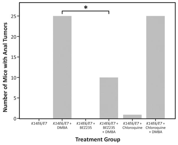
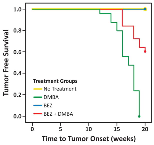
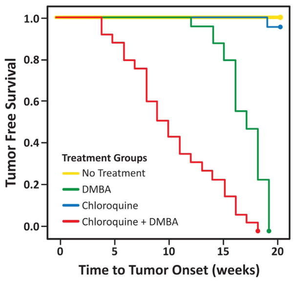
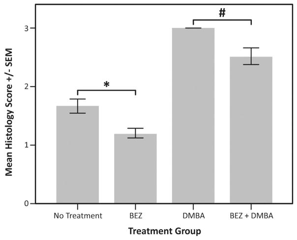
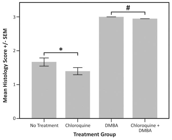
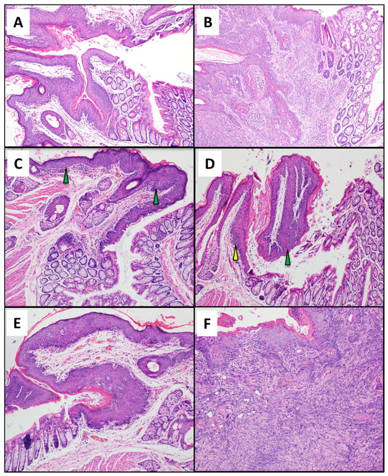
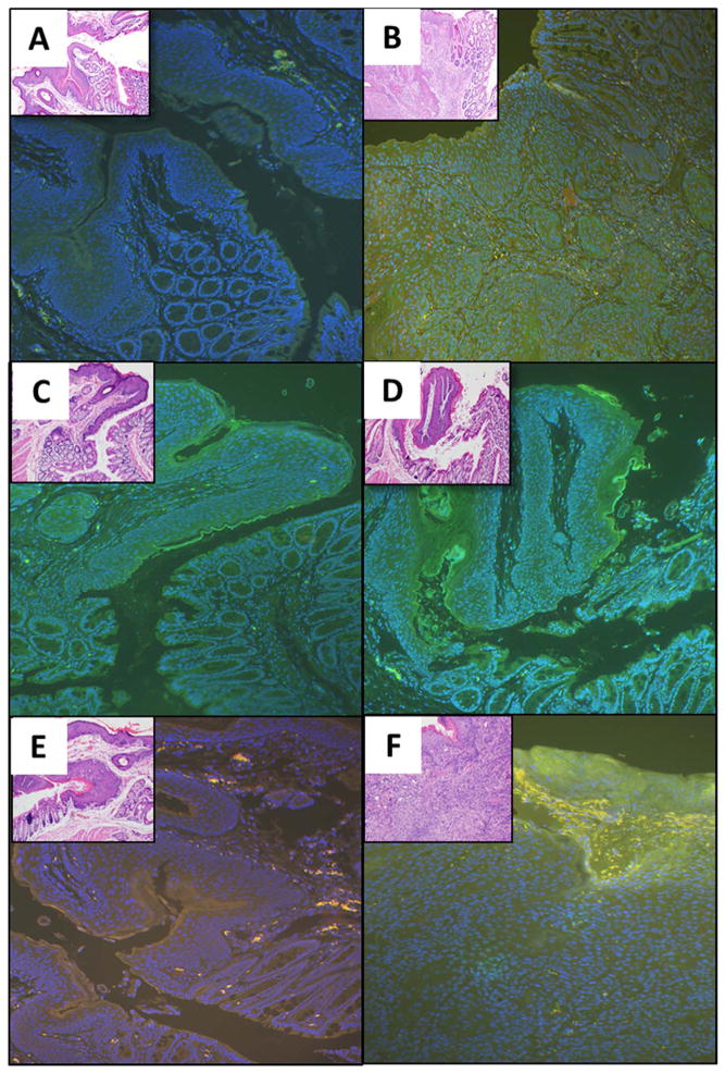
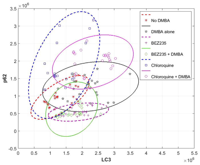
Autophagy is an intracellular, catabolic process that maintains cellular health. We examined the response of pharmacologic modulation of autophagy in an HPV mouse model of anal carcinogenesis. K14E6/E7 mice were treated with the topical carcinogen DMBA weekly and assessed for tumors over 20 weeks. Concurrently, they were given either chloroquine or BEZ235, to inhibit or induce autophagy, respectively. Time to tumor onset was examined. Immunofluorescence (IF) was performed for LC3β and p62 to examine autophagy. All DMBA treated K14E6/E7 mice developed anal cancer, contrary to zero of the no DMBA treated mice. Chloroquine plus DMBA resulted in a significant decrease in the time to tumor onset compared to K14E6/E7 treated with DMBA. Only 40% BEZ235 plus DMBA treated mice developed anal cancer. Autophagic induction with DMBA and BEZ235, and autophagic inhibition with chloroquine were confirmed via IF. Anal carcinogenesis can be inhibited or induced via pharmacologic modulation of autophagy.

Keywords: Anal cancer; Anal dysplasia; Autophagy; BEZ235; Chemoprevention; HPV.


Figures

Fig. 1
Fig. 1. Tumor incidence in treatment groups
100% of DMBA-only K14E6/E7 mice (25 mice) had anal tumors by the end of the 20-week period, while none of the no DMBA treated mice developed anal tumors. When treated with NVP-BEZ235 and DMBA concurrently, only 10 of the 25 K14E6/E7 mice showed signs of anal tumor development over the 20-week DMBA time course. Fifteen (60%) of the mice did not experience any anal tumor growth by the end of the 20-week DMBA treatment period (*P=0.001). There was no difference in the mice treated with chloroquine compared to the K14E6/E7 mice with and without DMBA.
Fig. 2
Fig. 2. Tumor free survival of mice treated with an autophagic inducer (BEZ235)
On average, K14E6/E7 mice receiving BEZ235 in the setting of DMBA (red had longer tumor free survival than those receiving DMBA alone (green, P < 0.0005). None of the mice in BEZ alone (blue) and no treatment control (yellow) groups developed tumors by 20 weeks. (For interpretation of the references to color in this figure legend, the reader is referred to the web version of this article.)
Fig. 3
Fig. 3. Tumor free survival of mice treated with an autophagic inhibitor (chloroquine)
On average, K14E6/E7 mice receiving chloroquine in the setting of DMBA (red had significantly shorter tumor free survival than those receiving DMBA alone (green, P < 1e−7). One of the mice in chloroquine alone group (blue) and none of the no treatment control (yellow) group mice developed tumors by 20 weeks. (For interpretation of the references to color in this figure legend, the reader is referred to the web version of this article.)
Fig. 4
Fig. 4. Average histologic score in mice treated with an autophagic inducer (BEZ235)
At the time of death or 20 weeks, H & E stained tissue from each mouse was graded histologically. The following scoring method was applied: normal histology =0, low-grade dysplasia =1, high-grade dysplasia =2, squamous cell carcinoma (any grade) =3. The average score for mice in each treatment group was then compared using a one-way ANOVA. Those mice receiving DMBA with BEZ235 (average score ± SD, 2.5 ± 0.7) or without BEZ235 (3 ± 0) were significantly different (#P=0.005). Additionally, untreated mice (1.7 ± 0.6) and those receiving BEZ235 (1.2 ± 0.4) alone were significantly different (*P=0.008).
Fig. 5
Fig. 5. Average histologic score in mice treated with an autophagic inhibitor (chloroquine)
At the time of death or 20 weeks, H & E stained tissue from each mouse was graded histologically. The following scoring method was applied: normal histology =0, low-grade dysplasia =1, high-grade dysplasia =2, squamous cell carcinoma (any grade) =3. The average score for mice in each treatment group was then compared using a one-way ANOVA. Those mice receiving DMBA with chloroquine (average score ± SD, 3 ± 0) or without chloroquine (3 ± 0) were not significantly different (#P=1.0). Additionally, untreated mice (1.7 ± 0.6) and those receiving chloroquine (1.4 ± 0.5) alone were not significantly different (*P=0.07).
Fig. 6
Fig. 6. Histological analysis of the anal transition zone for various treatment groups
Anal histology based on the findings identified by a trained pathologist. Anal tissue was examined at 20 weeks for each of the treatment groups. Magnification of each image is 200x. (A) K14E6/E7- low-grade dysplasia; (B) K14E6/E7 with DMBA - Grade 1 squamous cell carcinoma; (C) K14E6/E7 with BEZ235 - low-grade dysplasia with focal high-grade dysplasia; (D) K14E6/E7 with BEZ235 and DMBA - high-grade dysplasia; (E) K14E6/E7 with chloroquine - high-grade dysplasia; and (F) K14E6/E7 with chloroquine and DMBA - grade 3 squamous cell carcinoma.
Fig. 7
Fig. 7. Immunofluorescence for autophagic proteins following pharmacologic modulation of autophagy (magnification 200x)
Immunofluorescence was performed for autophagic protein LC3β (green) and autophagy-specific substrate p62 (red). DAPI (blue) was utilized as a nuclear counterstain. Anal tissue was examined at 20 weeks for each of the treatment groups: (A) K14E6/E7 (low-grade dysplasia); (B) K14E6/E7 with DMBA (Grade 1 squamous cell carcinoma); (C) K14E6/E7 with BEZ235 (high-grade dysplasia); (D) K14E6/E7 with BEZ235 and DMBA (low-grade dysplasia with focal high-grade dysplasia); (E) K14E6/E7 with chloroquine (high-grade dysplasia); and (F) K14E6/E7 with chloroquine and DMBA (Grade 3 squamous cell carcinoma). (For interpretation of the references to color in this figure legend, the reader is referred to the web version of this article.)
Fig. 8
Fig. 8. LC3β and p62 immunofluorescence levels in K14E6/E7 mice for various treatment groups as determined by FIJI analysis
Dual immunoflourescent (LC3β and p62) staining and high magnification imaging (20x) was performed. LC3β immunoflourescent intensity, indicating autophagic induction, is displayed on the x-axis and p62 immunoflourescent intensity, indicating autophagic function, on the y-axis. Ellipses represent the 95% confidence interval for each treatment group in the corresponding color. Solid lines indicate those groups that also received DMBA. One-way ANOVA analysis demonstrated a significant increase in LC3β in K14E6/E7 mice treated with DMBA versus no DMBA treated mice (P < 0.001). There was also a significant increase in LC3β in mice treated with BEZ235 alone compared to no DMBA treated mice (P < 0.001). In those mice treated with chloroquine, there was no change in LC3β in mice treated with chloroquine alone compared to no DMBA treated mice (P=0.92). In terms of p62 levels there was a statistically significant decrease in mice treated with BEZ235 with and without DMBA, compared to DMBA and no DMBA alone treated mice, and mice treated with DMBA and no DMBA with chloroquine (P=0.011 for BEZ235 with DMBA versus no DMBA; P < 0.001 for all other comparisons). There was also a statistically significant increase in p62 in chloroquine treated mice (with and without DMBA) compared to K14E6/E7 mice without DMBA, and with and without BEZ235 (P < 0.001 for all comparisons).

Similar articles

Cited by

References

    1. Barth S, Glick D, Macleod KF. Autophagy: assays and artifacts. J Pathol. 2010;221:117–124. - PMC - PubMed
    1. Carchman EH, Matkowskyj KA, Meske L, Lambert PF. Dysregulation of autophagy contributes to anal carcinogenesis. PLoS One. 2016;11:e0164273. - PMC - PubMed
    1. Chin-Hong PV. Human Papillomavirus in kidney transplant recipients. Semin Nephrol. 2016;36:397–404. - PMC - PubMed
    1. Glick D, Barth S, Macleod KF. Autophagy: cellular and molecular mechanisms. J Pathol. 2010;221:3–12. - PMC - PubMed
    1. Griffin LM, Cicchini L, Pyeon D. Human papillomavirus infection is inhibited by host autophagy in primary human keratinocytes. Virology. 2013;437:12–19. - PMC - PubMed

Publication types

MeSH terms