. 2017 Sep:74:110-124.
doi: 10.1016/j.dci.2017.04.009. Epub 2017 Apr 19.

Hemolymph proteins of Anopheles gambiae larvae infected by Escherichia coli

Affiliations

Hemolymph proteins of Anopheles gambiae larvae infected by Escherichia coli

Xuesong He et al. Dev Comp Immunol. 2017 Sep.

Abstract

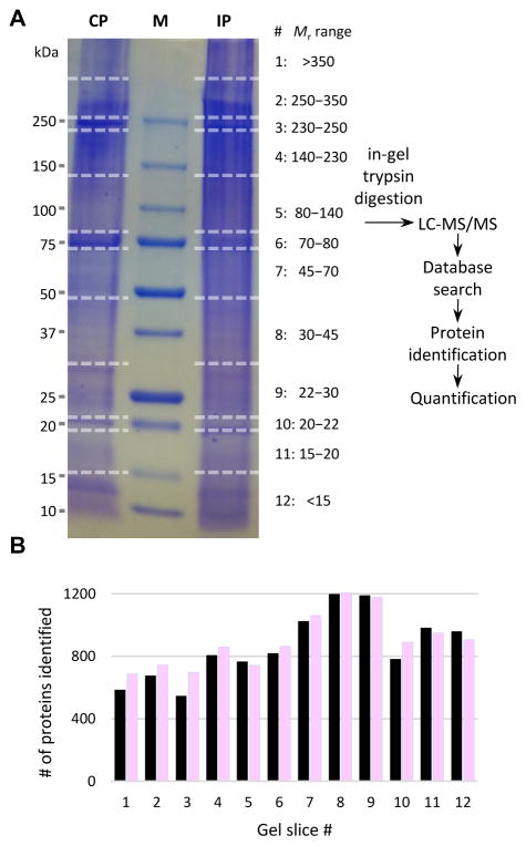
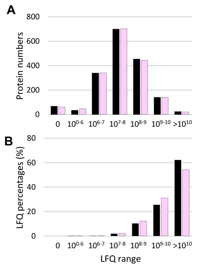
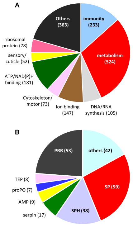
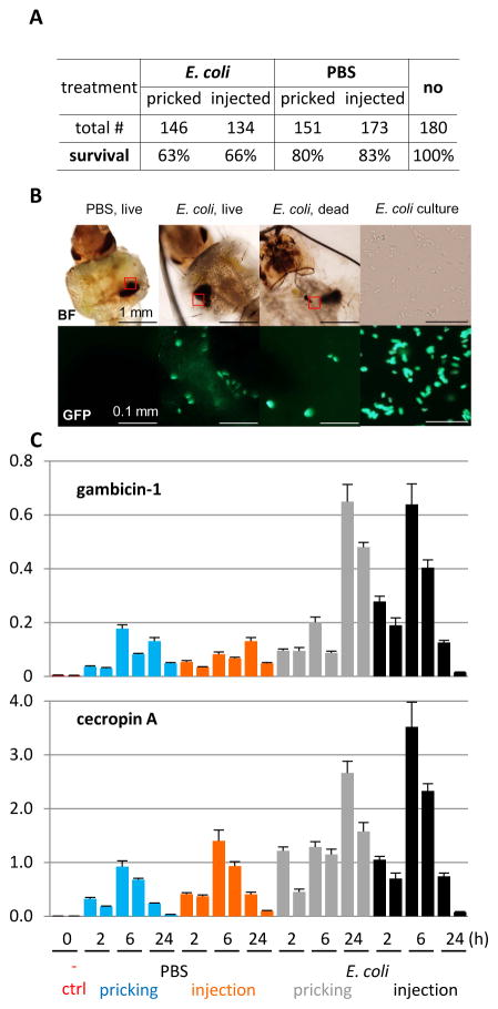
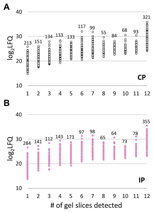
Anopheles gambiae is a major vector of human malaria and its immune system in part determines the fate of ingested parasites. Proteins, hemocytes and fat body in hemolymph are critical components of this system, mediating both humoral and cellular defenses. Here we assessed differences in the hemolymph proteomes of water- and E. coli-pricked mosquito larvae by a gel-LC-MS approach. Among the 1756 proteins identified, 603 contained a signal peptide but accounted for two-third of the total protein amount on the quantitative basis. The sequence homology search indicated that 233 of the 1756 may be related to defense. In general, we did not detect substantial differences between the control and induced plasma samples in terms of protein numbers or levels. Protein distributions in the gel slices suggested post-translational modifications (e.g. proteolysis) and formation of serpin-protease complexes and high Mr immune complexes. Based on the twenty-five most abundant proteins, we further suggest that major functions of the larval hemolymph are storage, transport, and immunity. In summary, this study provided first data on constitution, levels, and possible functions of hemolymph proteins in the mosquito larvae, reflecting complex changes occurring in the fight against E. coli infection.

Keywords: Gel mobility shift; Hemolymph proteins; Insect immunity; LC-MS/MS; Label-free quantification; Serine protease.


Figures

Fig. 1
Fig. 1. Schematic overview of the sample treatment and data analysis
(A) Hemolymph from the larvae challenged with E. coli and water was separately pooled (20–25 insects per group) to generate four induced (IP) and four control (CP) plasma samples (see Materials and Methods). The eight plasma samples were separated by SDS-PAGE on a 4–15% gradient gel that was later cut into 96 pieces. After in-gel trypsinolysis, peptides were separately extracted and analyzed by LC-MS/MS. Protein identification and quantification were performed as described in Materials and Methods. Sizes and positions of the Mr markers (lane M) are labeled on the left, while gel slice numbers (#’s) and corresponding Mr ranges of the gel slices are indicated on the right. (B) Distributions of the number of proteins identified in each gel slice. Black = CP; pink = IP.
Fig. 2
Fig. 2. Categorization of the total (1,756) and putative defense (233) proteins
(A) The 1,756 proteins classified into nine arbitrary groups with their names and number of proteins in each group indicated. (B) The 233 defense proteins were subdivided into seven groups: pattern recognition receptor (PRR), serine protease (SP), serine protease homolog (SPH), serpin, antimicrobial protein (AMP), prophenoloxidase (proPO), thioester protein (TEP), and others, with their counts marked.
Fig. 3
Fig. 3. Distributions of protein numbers (A) and LFQ percentages (B) in various LFQ ranges
Within each range, sums of LFQs of the proteins identified in CP (black) and IP (pink) are used to calculate their percentages in the grand totals (ΣLFQCP and ΣLFQIP), respectively.
Fig. 4
Fig. 4. Effects of sterile and septic wounding and injection on A. gambiae larvae
(A) Survival statistics; (B) Localization of GFP-labeled bacteria in the living and dead larvae 24 h after pricking with PBS or E. coli. Blurring of the signals was caused by their movements during imaging. upper: bright field; lower: fluorescence. Due to movement of E. coli cells in the culture (right), their positions in the bright field no longer corresponded to those in the fluorescence microscope image obtained a few minutes later. (C) Expression profiles of gambicin-1 (upper) and cecropin A (lower) genes after PBS and E. coli pricking and injection. Two pools of naïve larvae (10 insect per group) were as used as negative controls. The AMP mRNA levels relative to A. gambiae rpS7 are shown for each sample as mean ± standard deviation (n = 3). Two biological replicates were analyzed for all these samples.
Fig. 5
Fig. 5. Relationships of protein abundances and number of the gel slices they were identified
Relative abundances, log2LFQ, for proteins identified in CP (open circles, panel A) or IP (pink dots, panel B) and numbers of gel slices they were detected were plotted. The number of proteins within each group is marked above the data series.

References

    1. Abraham EG, Pinto SB, Ghosh A, Vanlandingham DL, Budd A, Higgs S, Kafatos FC, Jacobs-Lorena M, Michel K. An immune-responsive serpin, SRPN6, mediates mosquito defense against malaria parasites. Proc Natl Acad Sci USA. 2005;102:16327–16332. - PMC - PubMed
    1. Aguilar R, Jedlicka AE, Mintz M, Mahairaki V, Scott AL, Dimopoulos G. Global gene expression analysis of Anopheles gambiae responses to microbial challenge. Insect Biochem Mol Biol. 2005;35:709–719. - PubMed
    1. Ahrné E, Molzahn L, Glatter T, Schmidt A. Critical assessment of proteome-wide label-free absolute abundance estimation strategies. Proteomics. 2013;13:2567–2578. - PubMed
    1. Amenya DA, Chou W, Li J, Yan G, Gershon PD, James AA, Marinotti O. Proteomics reveals novel components of the Anopheles gambiae eggshell. J Insect Physiol. 2010;56:1414–1419. - PMC - PubMed
    1. An C, Budd A, Kanost MR, Michel K. Characterization of a regulatory unit that controls melanization and affects longevity of mosquitoes. Cell Mol Life Sci. 2011;68:1929–1939. - PMC - PubMed

MeSH terms