. 2017 Jun 16;83(13):e00561-17.
doi: 10.1128/AEM.00561-17. Print 2017 Jul 1.

Genome-Wide Investigation of Biofilm Formation in Bacillus cereus

Affiliations

Genome-Wide Investigation of Biofilm Formation in Bacillus cereus

Fang Yan et al. Appl Environ Microbiol. .

Abstract

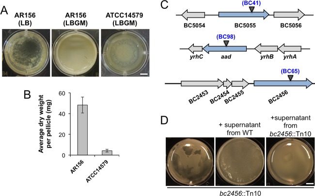
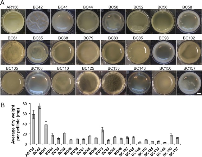
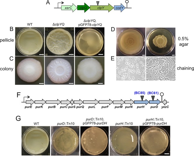
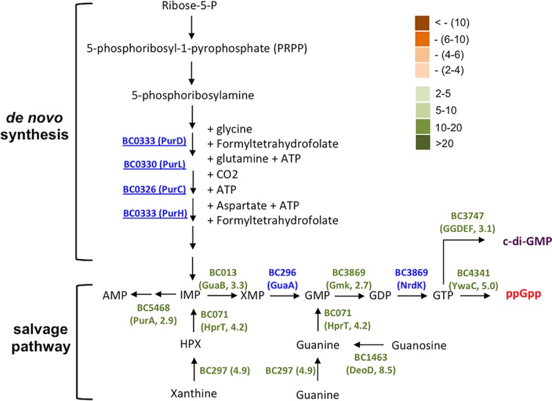
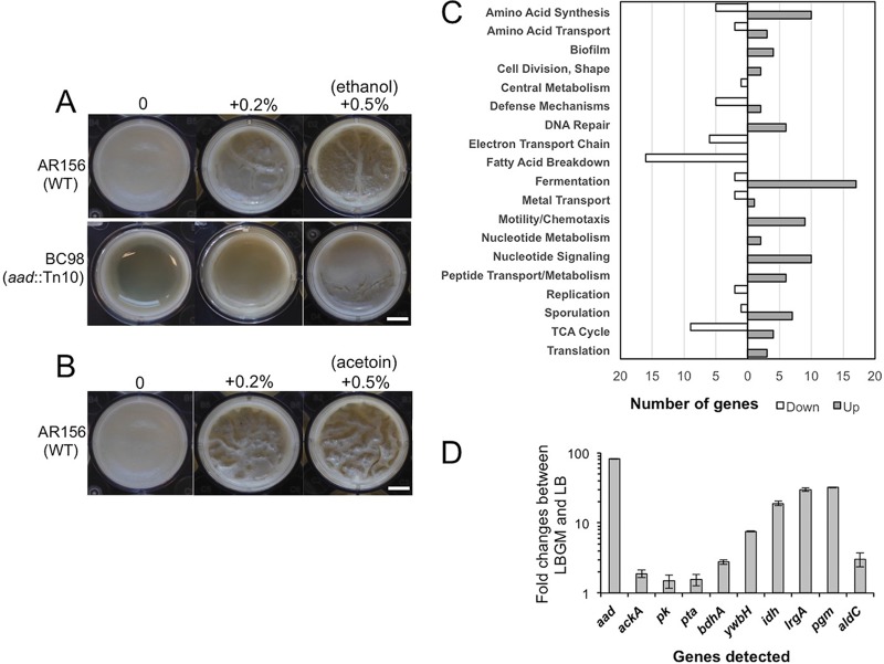
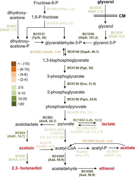
Bacillus cereus is a soil-dwelling Gram-positive bacterium capable of forming structured multicellular communities, or biofilms. However, the regulatory pathways controlling biofilm formation are less well understood in B. cereus In this work, we developed a method to study B. cereus biofilms formed at the air-liquid interface. We applied two genome-wide approaches, random transposon insertion mutagenesis to identify genes that are potentially important for biofilm formation, and transcriptome analyses by RNA sequencing (RNA-seq) to characterize genes that are differentially expressed in B. cereus when cells were grown in a biofilm-inducing medium. For the first approach, we identified 23 genes whose disruption by transposon insertion led to altered biofilm phenotypes. Based on the predicted function, they included genes involved in processes such as nucleotide biosynthesis, iron salvage, and antibiotic production, as well as genes encoding an ATP-dependent protease and transcription regulators. Transcriptome analyses identified about 500 genes that were differentially expressed in cells grown under biofilm-inducing conditions. One particular set of those genes may contribute to major metabolic shifts, leading to elevated production of small volatile molecules. Selected volatile molecules were shown to stimulate robust biofilm formation in B. cereus Our studies represent a genome-wide investigation of B. cereus biofilm formation.IMPORTANCE In this work, we established a robust method for B. cereus biofilm studies and applied two genome-wide approaches, transposon insertion mutagenesis and transcriptome analyses by RNA-seq, to identify genes and pathways that are potentially important for biofilm formation in B. cereus We discovered dozens of genes and two major metabolic shifts that seem to be important for biofilm formation in B. cereus Our study represents a genome-wide investigation on B. cereus biofilm formation.

Keywords: Bacillus cereus; biofilm formation; transcriptome; transposon mutagenesis.


Figures

FIG 1
FIG 1
B. cereus AR156 formed robust pellicle biofilms in the biofilm-inducing medium LBGM. (A) Pellicle biofilm formation by B. cereus AR156 in LB and LBGM, and by B. cereus ATCC 14579 in LBGM. Scale bar, 5 mm. (B) The average dry weights of the individual pellicle biofilms formed by AR156 and ATCC 14579 in LBGM were assayed by using the method described in Materials and Methods. The dry weight was calculated as milligrams per pellicle biofilm and was averaged from at least 3 independent samples. Error bars represent standard deviations. (C) The transposon insertion sites within specific genes on the chromosome of AR156 on selected mutants (those discussed in the Results) are indicated by triangles. The corresponding transposon insertion mutant was also indicated. Annotation of the genes is based on either the NCBI database entry for the sequenced B. cereus ATCC 14579 genome, or in some cases, on a BLAST search against strong homologous genes in the closely related B. subtilis. (D) Pellicle biofilm formation by the transposon insertion mutant BC65 (bc2456::Tn10) in LBGM, and in LBGM supplemented with the concentrated cell-free supernatant from the wild-type AR156 or that from the transposon insertion mutant BC65. Scale bar, 5 mm.
FIG 2
FIG 2
Pellicle biofilm formation by 23 transposon insertion mutants. (A) Twenty-three individual transposon insertion mutants and the wild-type AR156 were assayed for pellicle biofilm formation in LBGM. Scale bar, 3 mm. (B) The average dry weights of the individual pellicle biofilms formed by the mutants and AR156 were assayed. The dry weight was calculated as milligram per pellicle biofilm and was averaged from at least 3 independent samples. Error bars represent standard deviations.
FIG 3
FIG 3
(A to E) The ΔclpYQ deletion mutant formed more robust biofilms while impaired for swarming motility and cell separation. (A) A schematic drawing of the presumptive operon of xerC-clpY-clpQ-codY in B. cereus. The transposon insertion site within the clpY gene in BC42 is indicated by the triangle. (B and C) Pellicle (B) and colony (C) biofilms formed by wild-type AR156 and the clpYQ in-frame deletion mutant (FY178), and the clpYQ complementation strain (YY250) in LBGM. Scale bars, 5 mm (B) and 2 mm (C). (D) Swarming motility of AR156 and FY178 on LB plates solidified with 0.5% agar. Plates were incubated at 30°C for 12 h before images were taken. Scale bars represent 1 cm. (E) Cells of AR156 and FY178 grown to late-exponential phase (OD600, 1) and observed under bright-field microscopy. Scale bars, 10 μm. (F and G) Genes involved in purine biosynthesis are important for biofilm formation in B. cereus. (F) A schematic drawing of the putative purine biosynthesis gene cluster in B. cereus AR156. The transposon insertion sites within the purH (BC85) and purD (BC61) genes are indicated by triangles. (G) Pellicle biofilm formation in LBGM by AR156, the two transposon insertion mutants, BC61 (purD::Tn) and BC85 (ΔpurH::Tn), and the two complementation strains, YY251(purD::Tn, pGFP78-purDH) and YY252(ΔpurH::Tn, pGFP78-purDH). Scale bars, 5 mm.
FIG 4
FIG 4
Genes in the proposed pathway for purine biosynthesis and GTP homeostasis were strongly upregulated during biofilm induction in B. cereus. Genes which are involved in the proposed pathway and whose expression was seen to be significantly altered under biofilm induction (in the RNA-seq experiment) are annotated. The fold change in gene expression was also indicated. Significantly increased expression (from 2- to >20-fold) under biofilm induction was seen for genes in green. Significantly decreased expression (from 2- to >10-fold) was seen for genes in orange. Genes in blue indicate insignificant change (<2-fold) in gene expression. Numbers next to the gene name in parentheses represent the fold changes in gene expression.
FIG 5
FIG 5
(A and B) Ethanol- and acetoin-stimulated biofilm formation in B. cereus. (A) Pellicle biofilm formation by AR156 and BC98(aad::Tn) in LBGM in the presence or absence of ethanol. (B) Pellicle biofilm formation by AR156 in LBGM in the presence or absence of acetoin. Scale bars, 2.5 μm. WT, wild type. (C and D) Genome-wide transcriptome analyses of B. cereus AR156 cells during biofilm induction. (C) Summary of the genome-wide transcriptome analysis by RNA-seq. A total of about 500 genes whose expression was significantly altered under biofilm induction (in LBGM) compared to non-biofilm induction (in LB), are categorized into differential functional groups. The number of genes in each functional group was calculated based on the predicted function of the genes. Numbers on the x axis represent number of genes showing differential expression. (D) Validation of upregulation of 10 selected genes under biofilm induction (in LBGM) compared to non-biofilm induction (in LB) by reverse transcription-quantitative PCR (RT-qPCR). Down, downregulated; Up, upregulated.
FIG 6
FIG 6
The proposed metabolic shift during biofilm induction in B. cereus. Most genes involved in this predicted metabolic pathway were found to be highly upregulated under biofilm induction (in LBGM) compared to non-biofilm induction (in LB) based on the results of the genome-wide transcriptome analysis by RNA-seq. The genes with different colors represent the difference in fold change in differential expression (LBGM versus LB). Dark green stands for highest induction, while dark brown stands for strongest repression. Numbers next to the gene name in the parentheses represent the fold changes in gene expression.

Similar articles

Cited by

References

    1. O'Toole G, Kaplan HB, Kolter R. 2000. Biofilm formation as microbial development. Annu Rev Microbiol 54:49–79. doi:10.1146/annurev.micro.54.1.49. - DOI - PubMed
    1. Stoodley P, Sauer K, Davies DG, Costerton JW. 2002. Biofilms as complex differentiated communities. Annu Rev Microbiol 56:187–209. doi:10.1146/annurev.micro.56.012302.160705. - DOI - PubMed
    1. Hall-Stoodley L, Stoodley P. 2009. Evolving concepts in biofilm infections. Cell Microbiol 11:1034–1043. doi:10.1111/j.1462-5822.2009.01323.x. - DOI - PubMed
    1. Emmert EAB, Handelsman J. 1999. Biocontrol of plant disease: a (Gram-) positive perspective. FEMS Microbiol Lett 171:1–9. doi:10.1111/j.1574-6968.1999.tb13405.x. - DOI - PubMed
    1. Chen Y, Yan F, Chai Y, Liu H, Kolter R, Losick R, Guo J-h. 2013. Biocontrol of tomato wilt disease by Bacillus subtilis isolates from natural environments depends on conserved genes mediating biofilm formation. Environ Microbiol 15:848–864. doi:10.1111/j.1462-2920.2012.02860.x. - DOI - PMC - PubMed

Publication types

Substances

LinkOut - more resources