. 2017 Jun 15;6(6):765-776.
doi: 10.1242/bio.024273.

Centrin diversity and basal body patterning across evolution: new insights from Paramecium

Affiliations

Centrin diversity and basal body patterning across evolution: new insights from Paramecium

Anne Aubusson-Fleury et al. Biol Open. .

Abstract

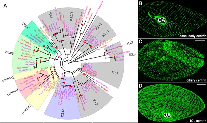
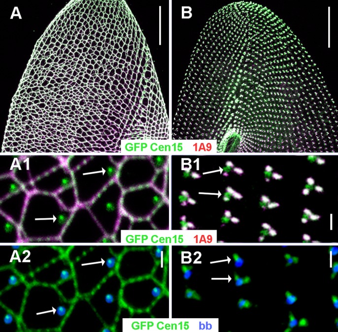
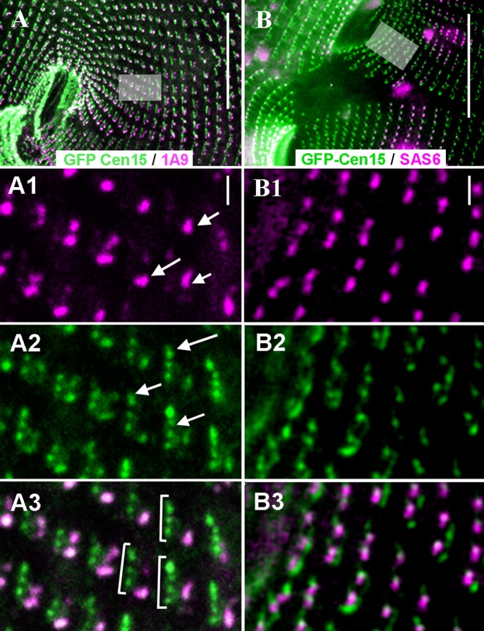
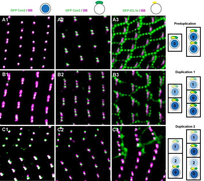
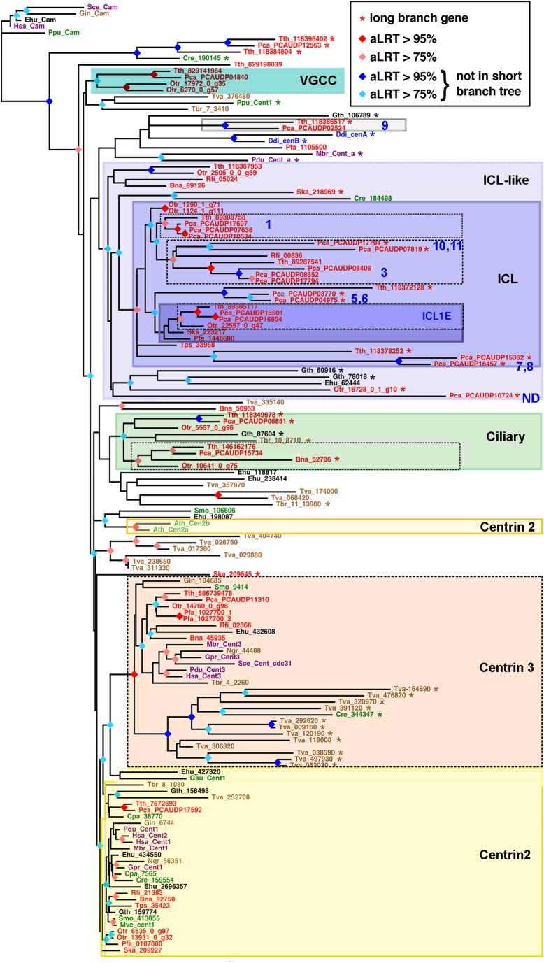
First discovered in unicellular eukaryotes, centrins play crucial roles in basal body duplication and anchoring mechanisms. While the evolutionary status of the founding members of the family, Centrin2/Vfl2 and Centrin3/cdc31 has long been investigated, the evolutionary origin of other members of the family has received less attention. Using a phylogeny of ciliate centrins, we identify two other centrin families, the ciliary centrins and the centrins present in the contractile filaments (ICL centrins). In this paper, we carry on the functional analysis of still not well-known centrins, the ICL1e subfamily identified in Paramecium, and show their requirement for correct basal body anchoring through interactions with Centrin2 and Centrin3. Using Paramecium as well as a eukaryote-wide sampling of centrins from completely sequenced genomes, we revisited the evolutionary story of centrins. Their phylogeny shows that the centrins associated with the ciliate contractile filaments are widespread in eukaryotic lineages and could be as ancient as Centrin2 and Centrin3.

Keywords: Basal body anchoring; Basal body assembly; Centrin evolution; Ciliary centrins; Ciliated epithelia polarity.


Conflict of interest statement

Competing interestsThe authors declare no competing or financial interests.

Figures

Fig. 1.
Fig. 1.
Ciliate centrins: phylogeny and localisation in Paramecium. (A) Maximum-likelihood unrooted tree of ciliate centrins. The robustness of the nodes is coded by coloured diamonds: pink for aLRT scores superior to 75%, red for aLRT scores superior to 95%. Boxes indicate well supported sub-groups, from either the phylogenetical or functional point of view. Pte (purple): Paramecium tetraurelia; Pca (pink): Paramecium caudatum; Tth (red): Tetrahymena thermophila; Otr (brown): Oxytricha trifallax. Two outgroup sequences for Centrin2 and Centrin3 families have been added: Cre/VFL2 (green), Chlamydomonas reinhardti and Sce/cdc31 (grey), Saccharomyce cerevisiae. (B-D) Confocal images. GFP-tagging of representatives of these diverse centrins reveals their three major localisations: at basal bodies (B), in cilia (C) and along the infraciliary lattice (D) labelled by GFP-Centrin2, GFP-Cent14 and GFP-ICL1e, respectively. OA, oral apparatus. Scale bar: 20 µm.
Fig. 2.
Fig. 2.
Wild-type and ICL-less (BP) cells: respective localisations of ICL1e, ICL centrins and basal bodies. Confocal images. (A) Wild-type cell triple-labelled by GFP-ICL1e (green), the anti-ICL 1A9 (magenta) and the anti-tubulin PolyE to label basal bodies (blue): ventral side showing the ICL network. Insets: the GFP-labelling shows a dotted pattern along the meshes of the network (A1) and a singular dot anterior right to each basal body (A2). (B) Two BP cells double-labelled with GFP-ICL1e (green) and 1A9 (B and B1) and with GFP-ICL1e and the PolyE (B2). (B1) remnants of the ICL network are labelled by both 1A9 and GFP-ICL1e, except for a dot which is not decorated by 1A9. (B2) The GFP-ICL1e dot is detected close to each basal body as in the wild type. Arrows: ICL1e dot. Scale bar: 20 µm in A, B; 1 µm in A1-2, B1-2.
Fig. 3.
Fig. 3.
The concerted duplication of basal bodies and ICL1e complexes in ICL-less BP cells. Epifluorescence images. (A-A3) Sequential assembly of the ICL1e complex and other ICL components. At the beginning of division, new basal bodies are assembled anterior to parental ones, resulting in a series of closely juxtaposed basal bodies (see Fig. S2A). In this dividing BP cell (A) expressing GFP-Cen15 (green) and labelled by 1A9 (magenta), basal body duplication has already begun resulting in series (brackets in A3) of new ICL1e complexes (arrows in A2) associated with just duplicated basal bodies. By contrast, the 1A9 labelling (arrows in A1) is only associated with the parental (posterior) basal bodies of each series. (B-B3) Respective timings of duplication of basal bodies and ICL1e dots. In this cell co-expressing GFP-Cen15 (green) and RFP-SAS6 (magenta), on both sides of the fission furrow, an ICL1e complex flanks anteriorly each SAS-6 dot (magenta) marking all basal bodies, whether pre-existing or just formed. Scale bars: 20 µm in A, B; 1 µm in A1-3, B1-3.
Fig. 4.
Fig. 4.
Effects of ICL1e inactivation on the basal body pattern. Epifluorescence images. (A) In wild-type cells, by the first/second division upon inactivation, the ICL network (magenta) is disrupted and mis-positioned basal bodies (1D5, green) are detected. Scale bar: 20 µm; inset: ×3.5. (B) In BP cells, by the first /second division upon inactivation, basal bodies without associated ICL remnants (magenta) are detected (1D5 green, arrows). Scale bar: 20 µm; inset: ×3. Insets in A and B are outlined by grey squares in corresponding figures. (C) Numerous internal basal bodies are detected (arrows). Scale bar: 20 µm. (D,E) Transmission electron microscopy of ICL1e-inactivated wild-type cell. The unanchored basal bodies appear fully developed with a transition zone (large arrow) and associated ring structure (short arrows). Scale bar: 200 nm.
Fig. 5.
Fig. 5.
Topological relationships between Centrin2, Centrin3 and ICL1e during basal body duplication. Confocal images. Three successive stages of basal body duplication are illustrated in cells expressing GFP-Centrin2 (A1-C1), GFP-Centrin3 (A2-C2) or GFP-ICL1e (A3-C3) and labelled with the ID5 antibody. (A) Pre-duplication, (B) duplication 1 leading to one basal body per cortical units, and (C) duplication 2, resulting in paired basal bodies in some units. The scheme to the right summarises the specific behaviour of each of the three centrins at successive stages. Circles: basal bodies; colour intensities represent successive basal bodies generations (0, parental basal bodies; 1, basal bodies from duplication 1; 2, basal bodies from duplication 2); blue, Centrin2; green, Centrin3; yellow, ICL1e. While the Centrin2 labelling (A and blue) is superimposed to the basal bodies at all three stages, Centrin3 (2 and green) and ICL1e (3 and yellow) all localise anterior of all basal bodies during the pre-duplication stage (A), and anterior of the single and of the sole posterior basal body in paired basal bodies at other stages (B and C). Each image represents a 10×10 µm area.
Fig. 6.
Fig. 6.
Localisation of GFP-ICL1e upon inactivation of Centrin2 and Centrin3. Confocal images. Green, GFP-ICL1e; magenta, basal bodies labelled with ID5. Insets are indicated by grey squares. (A) Upon inactivation of Centrin2, intra-cytoplasmic basal bodies are devoid of the ICL1e labelling. (B) In contrast, upon inactivation of Centrin3, intra-cytoplasmic basal bodies display the ICL1e labelling. In both cases, ICL remnants are detected at the cell surface (large arrows). Thin arrows, intracytoplasmic microtubules. OA, oral apparatus. Scale bar: 20 µm; ×2 in insets.
Fig. 7.
Fig. 7.
Localisation of GFP-Centrin3 upon inactivation of ICL1e. (A-D) Confocal and (E-H) STED images. Cells expressing GFP-Centrin3 (green) were immunolabelled with ID5 (magenta) to decorate the basal bodies. (A,C,E,G) Control, (B,D,F,H) ICL1e-depleted cell. Scale bars: 20 µm. The areas shown in C and D are outlined in A and B. (A) Wild-type cell, Centrin3 is associated with all basal bodies. (C) All spots of Centrin3 associated with single or paired basal bodies display a similar intensity. By the anterior basal body of the pairs, an additional faint spot is detected (arrows in C) suggesting a pre-duplication stage. Inset in C, lateral view of a basal body by STED imaging; the base and tip localisations (arrow) of the Centrin3 are detected. (E) Non-deconvoluted and (G) deconvoluted STED images of another Centrin3-expressing cell. The part magnified in the insets is outlined with an ellipse. Two Centrin3 spots anterior to single basal bodies and posterior basal body of paired ones, as well as an additional anterior spot associated with the anterior basal body of each pair are detected. Inset in E, the three spots are surrounded by a faint cloud of GFP-material, which could correspond to the GFP-Centrin3 at the tip of the basal body. After deconvolution (G), the three spots are clearly identified, but the surrounding cloud is no longer detected. Scale bars: 0.5 µm. (B) ICL1e-depleted cell. Basal bodies are less regularly aligned, and extra basal bodies are observed (arrows). (D) An alternance of strong (long arrows) and weak (short arrows) fluorescence of the GFP-Centrin3 spots associated with the basal bodies is observed along the antero-posterior rows. Some basal bodies have almost lost their Centrin3 (circles). Inset in D, lateral view of a basal body by STED imaging: the double (at the base and the tip) localisation (arrow) of the Centrin3 is detected but with a variable intensity depending upon the basal body. (F) Non-deconvoluted and (H) deconvoluted STED images of another cell. Left insets are outlined by a circle and right insets by an ellipse. After ICL1e depletion, the number and size of the GFP-Centrin3 spots are smaller. The right anterior Centrin3 spot is lost in most basal bodies, and some basal bodies have no spot at all while a tiny cloud of GFP-Centrin3 is still detected (F, left inset).
Fig. 8.
Fig. 8.
Maximum-likelihood tree of centrins. The colours of the gene labels indicate the lineage of origin (red, SAR; brown, Excavates; green, Archaeplastids; blue, Amoebozoans; purple, Opisthokonts). The ICL subfamilies of Paramecium are noted in blue numbers. ND, not determined. Species abbreviations: Ath, Arabidopsis thaliana; Bna, Bigelowiella natans; Cpa, Cyanophora paradoxa; Cre, Chlamydomonas reinhardtii; Ddi, Dictyostelium discoideum; Ehu, Emiliana huxleyi; Fi, Retyculomyxa filosa; Gin, Giardia intestinalis; Gpr, Gonapodya prolifera; Gsu, Garderia sulphuraria; Gth, Guillardia theta; Hsa, Homo sapiens; Mbr, Monosiga brevicolis; Mve, Marsilea vestita; Ngr, Naegleria gruberi; Otr, Oxytricha trifallax; Pca, Paramecium caudatum; Pdu, Platynereis dumerii; Pfa, Plasmodium falciparum 3D7; Ppu, Porphyridium purpureum; Sce, Saccharomyces cerevisiae; Ska, Symbiodinium kawagutii; Smo, Selaginella moellendorffii; Tbr, Trypanosoma brucei; Tps, Thalassiosira pseudonana; Tth, Tetrahymena thermophila; Tva, Trichomonas vaginalis. Stippled black boxes indicate well supported phylogenetic groups (red and orange nodes): Centrin3, Ciliate ciliary centrins, and ICL centrins corresponding to the P. tetraurelia ICL1, ICL3/10/11 and ICL1e centrins. Potential extension of these well supported groups with reduced tree support (blue nodes) are indicated in colours (blue for ICL/ICL-like and green for ciliary centrins). The group of Centrin2 sequences is only weakly supported and some sequences jump out of this group. This is for example the case in Arabidopsis in which the two centrin genes, not considered as Centrin2 by some authors (Hodges et al., 2011), nevertheless display some of its functional properties (Molinier et al., 2004; Nishi et al., 2013).

Similar articles

Cited by

References

    1. Adl S. M., Simpson A. G., Lane C. E., Lukeš J., Bass D., Bowser S. S., Brown M. W., Burki F., Dunthorn M., Hampl V. et al. (2012). The revised classification of eukaryotes. J. Eukaryot. Microbiol. 59, 429-493. 10.1111/j.1550-7408.2012.00644.x - DOI - PMC - PubMed
    1. Anisimova M. and Gascuel O. (2006). Approximate likelihood-ratio test for branches: a fast accurate and powerful alternative. Syst. Biol. 65, 639-652. 10.1080/10635150600755453 - DOI - PubMed
    1. Arnaiz O., Cain S., Cohen J. and Sperling L. (2007). ParameciumDB: a community resource that integrates the Paramecium tetraurelia genome sequence with genetic data. Nucleic Acids Res. 35, D439-D444. 10.1093/nar/gkl777 - DOI - PMC - PubMed
    1. Arnaiz O., Cohen J., Tassin A. M. and Koll F. (2014). Remodelling Cildb, a popular database for for cilia and links for ciliopathies. Cilia 3, 9 10.1186/2046-2530-3-9 - DOI - PMC - PubMed
    1. Aubusson-Fleury A., Lemullois M., Garreau de Loubresse N., Laligné C., Cohen J., Rosnet O., Jerka-Dziadosz M., Beisson J. and Koll F. (2012). The conserved centrosomal protein FOR20 is required for assembly of the transition zone and basal body docking at the cell surface. J. Cell Sci. 125, 4395-4404. 10.1242/jcs.108639 - DOI - PubMed

LinkOut - more resources