Comparative Study
. 2017 Jun 16;292(24):10048-10060.
doi: 10.1074/jbc.M116.771071. Epub 2017 Apr 21.

Dual regulation of Stat1 and Stat3 by the tumor suppressor protein PML contributes to interferon α-mediated inhibition of angiogenesis

Affiliations
Comparative Study

Dual regulation of Stat1 and Stat3 by the tumor suppressor protein PML contributes to interferon α-mediated inhibition of angiogenesis

Kuo-Sheng Hsu et al. J Biol Chem. .

Abstract

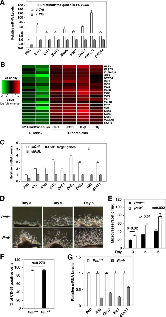
IFNs are effective in inhibiting angiogenesis in preclinical models and in treating several angioproliferative disorders. However, the detailed mechanisms of IFNα-mediated anti-angiogenesis are not completely understood. Stat1/2/3 and PML are IFNα downstream effectors and are pivotal regulators of angiogenesis. Here, we investigated PML's role in the regulation of Stat1/2/3 activity. In Pml knock-out (KO) mice, ablation of Pml largely reduces IFNα angiostatic ability in Matrigel plug assays. This suggested an essential role for PML in IFNα's anti-angiogenic function. We also demonstrated that PML shared a large cohort of regulatory genes with Stat1 and Stat3, indicating an important role of PML in regulating Stat1 and Stat3 activity. Using molecular tools and primary endothelial cells, we demonstrated that PML positively regulates Stat1 and Stat2 isgylation, a ubiquitination-like protein modification. Accordingly, manipulation of the isgylation system by knocking down USP18 altered IFNα-PML axis-mediated inhibition of endothelial cell migration and network formation. Furthermore, PML promotes turnover of nuclear Stat3, and knockdown of PML mitigates the effect of LLL12, a selective Stat3 inhibitor, on IFNα-mediated anti-angiogenic activity. Taken together, we elucidated an unappreciated mechanism in which PML, an IFNα-inducible effector, possess potent angiostatic activity, doing so in part by forming a positive feedforward loop with Stat1/2 and a negative feedback loop with Stat3. The interplay between PML, Stat1/Stat2, and Stat3 contributes to IFNα-mediated inhibition of angiogenesis, and disruption of this network results in aberrant IFNα signaling and altered angiostatic activity.

Keywords: STAT transcription factor; STAT3; angiogenesis; interferon; signal transducers and activators of transcription 1 (STAT1).


Conflict of interest statement

The authors declare that they have no conflicts of interest with the contents of this article

Figures

Figure 1.
Figure 1.
PML is required for IFNα-mediated angiostatic activity in vivo. Left, two representative Matrigel plugs from each group are shown. Right, quantification of angiogenesis. Approximately 200-mg plugs isolated from mice were homogenized in Drabkin's solution, and optical density was measured at 540 nm and normalized to the weight of the plugs. Unpaired two-tail t test (*, p < 0.05; ***, p < 0.001). The numbers (N) of mice are indicated for each group.
Figure 2.
Figure 2.
Depletion of PML promoted microvessel outgrowth and changed expression of IFNα and Stat1-inducible genes. A, qRT-PCR of IFNα-inducible genes in control and PML KD HUVECs. HUVECs were transiently transfected with a control siRNA or a mixture of two PML siRNAs. Total RNA was prepared 48 h post transfection followed by qRT-PCR using gene-specific primers. B, a heatmap representation of an array of genes that were significantly down-regulated in PML KD ECs (lanes 1 and 2) or significantly up-regulated in human fibroblasts (BJ cells) with overexpression of Stat1 (lane 3), U-Stat1 (lane 4), and treatment of IFNβ (lane 5) or IFNγ (lane 6) (data are from Cheon and Stark (37). The log-fold change was scaled in a green-dark-red color scheme. Each row represents a gene designated by the official gene symbol. Two independent PML siRNAs (siP1 and siP2) was compared with a non-targeting control siRNA (siC). C, RT-qPCR of U-Stat1 target genes in PML KD ECs. HUVECs were transiently transfected with a non-targeting siRNA or a mixture of siP-1 and siP-2. Total RNA was isolated 48 h after transfection followed by qRT-PCR. Relative mRNA levels are shown. D, representative pictures showing microvessel outgrowth in explanted aortic rings isolated from WT and Pml−/− mice at days 3, 5, and 6. Note that more microvessels sprouting from aortic rings prepared from Pml−/− mice than those isolated from the WT animals. E, quantification of microvessel outgrowth. F, cells from aortic ring outgrowth at the end of the experiments (12 days after explanting) were isolated and analyzed for CD31 expression by FACS analysis. G, aortic ECs were isolated from WT and Pml−/− mice as described in F. The expression of putative Stat1 target genes was quantified by RT-qPCR.
Figure 3.
Figure 3.
Long-term treatment of IFNα induced isgylation of Stat1 and Stat2. A, RT-qPCR of USP18 mRNA accumulation. B, effect of USP18 KD on Stat1 and Stat2 expression. HUVECs were transiently transfected with a control (siCtrl) or two independent siRNAs (siU-1 and siU-2) against USP18. 72 h after transfection cells were treated with vehicle or IFNα (103 units/ml) for 16 h. An aliquot of cells was used to isolate total RNA followed by RT-qPCR (A), and the rest of cells were used for subcellular fractionation (B). L.E., long exposure; S.E., short exposure.
Figure 4.
Figure 4.
The effect of USP18 or ISG15 knockdown on Stat1 and Stat2 isgylation in response to IFNα treatment. HUVECs were transfected with a control, ISG15, or USP18 siRNA. 72 h later cells were treated with vehicle or IFNα (103 units/ml) for 16 h and harvested, and subcellular fractions were prepared. The nuclear fractions were used for immunoprecipitation with a control IgG, anti-Stat1 (A), Isg15 (B and D), or anti-Stat2 (C) antibodies. Ten percent of the input and the immunopellets were Western-blotted with anti-Stat1 (A and B), anti-Isg15 (A and C), or anti-Stat2 (C and D) antibodies. The arrows mark isgylated Stat1 or Stat2 (A and C). The asterisks mark unmodified Stat1 or Stat2 (B and D, lane 7 and 8). Because Isg15-Stat1 and Isg15-Stat2 are in low abundance, only a longer exposure of the film was able to detect these two species in the Input (10%) (lanes 1–3, data not shown). S.E., shorter exposure; L.E., longer exposure.
Figure 5.
Figure 5.
The effects of IFNα on Stat1, Stat2, and Stat3 protein. HUVECs were transiently transfected with a control siRNA (siCtrl) or two independent USP18 siRNA (siU). 48 h after transfection, equal numbers of cells were seeded, treated with IFNα, and harvested at indicated times. Subcellular fractions were prepared, and nuclear fractions were subjected to Western blotting with anti-Stat1 (A), anti-Stat2 (B), and anti-Stat3 (C) antibodies. The arrows mark the putative isgylated Stat1 (A) and Stat2 (B). The asterisk marks the unmodified Stat3 (C). S.E., short exposure; L.E., longer exposure.
Figure 6.
Figure 6.
PML promoted Stat1 and Stat2 isgylation and affected ISGF3 complexes. A, HUVECs were transiently transfected with a control (siCtrl) or a PML siRNA (siPML). 72 h later cells were treated with IFNα, harvested at 0, 0.5, 1, 2, and 16 h, and subcellular fractions were prepared. Nuclear (lanes 1–10) and cytoplasmic (lanes 11–20) fractions were subjected to Western blotting with the indicated antibodies. L.E., long exposure; S.E., short exposure. The arrows indicate Isg15-nStat1 and Isg15-nStat2. The asterisk marks a unknown Stat1 species, likely to be Tyr(P)-701-Stat1. Lamin B and α-tubulin were used as markers for nuclear and cytoplasmic fractions, respectively. B, the effect of PML KD on the abundance of Tyr(P)-701-Stat1. Nuclear fractions from A were subjected to Western blotting with anti-Tyr(P)-701-Stat1 antibody. C, aortic ECs from Pml−/− show decreased Isg15-nStat1, Isg15-nStat2, and nIRF9. Aortic ECs isolated from aortic rings of WT and Pml−/− mice were treated with IFNα (103 units/ml) and harvested at the indicated times, and subcellular fractions were prepared. The nuclear fractions were subjected to Western blotting with the indicated antibodies.
Figure 7.
Figure 7.
The effect of USP18 knockdown on IFNα-mediated Stat1 isgylation, EC migration, and capillary tube formation. A, HUVECs were transiently transfected with a control (siCtrl), USP18 (siU), or PML (siPML) siRNAs for 48 h and trypsinized, and equal amounts of cells were seeded in 6-well plates. The levels of Stat1 and Stat1 isgylation were detected by Western blotting using antibodies against Stat1. Lamin B was used as a loading control. Arrows: isgylated Stat1. B, the left panel shows representative results of wound healing assays. The cell migration rates were quantified and are shown on the right panel. The statistical results are the mean ± S.D. (n = 6) (*, p < 0.05; ***, p < 0.001; unpaired two-tailed t test). C, PML and USP18 in HUVECs were knocked down by the indicated siRNAs. Cells were then treated with IFNα (103 units/ml) for 16 h followed by capillary tube formation assays. Representative images are shown on the left. The numbers of braches in each field were counted, and the quantitative results are shown in the right panel. The statistical results are mean ± S.D. (n = 6) (**, p < 0.01; ***, p < 0.001; unpaired two-tailed t test).
Figure 8.
Figure 8.
PML negatively regulated nStat3 stability and attenuated Stat3 function in EC network formation. A, accumulation of Tyr(P)-705-nStat3 in PML KD HUVECs. Procedures were the same as that described in Fig. 6, except that Stat3 and Tyr(P)-705-Stat3 antibodies were used for Western blotting. B, quantification of nStat3 protein abundance from A. The relative abundance of Stat3 at each time point was normalized to that at 0 h of IFNα treatment (lanes 1 and 6). n = 5. C, samples from A were subjected to Western blotting with anti-Tyr(P)-705-Stat3 antibody. D, MG132 increased nStat3 after 0.5 h of IFNα treatment. HUVECs were treated with 0.5 h of IFNα (103 units/ml) followed by 0.5 h of treatment with vehicle or MG132 and harvested, nuclear fractions were prepared and Western-blotted with the indicated antibodies. E, knockdown of PML abolished MG132-mediated accumulation of nStat3 in response to IFNα stimulation. F, control and PML KD HUVECs were pretreated with LLL12 (100 nm) for 3 h and trypsinized. Equal numbers of cells were subjected to wound-healing assays and capillary tube formation assays in the presence of IFNα (G). Statistical analysis was performed by counting numbers of branch points per field at 6 and 21 h of the assay. n = 6 per group (*, p < 0.05; ***, p < 0.001; unpaired two-tailed t test).

Similar articles

Cited by

References

    1. Gutterman J. U. (1994) Cytokine therapeutics: lessons from interferon α. Proc. Natl. Acad. Sci. U.S.A. 91, 1198–1205 - PMC - PubMed
    1. Borden E. C., Sen G. C., Uze G., Silverman R. H., Ransohoff R. M., Foster G. R., and Stark G. R. (2007) Interferons at age 50: past, current, and future impact on biomedicine. Nat. Rev. Drug Discov. 6, 975–990 - PMC - PubMed
    1. Liu F., Gao X., Wang J., Gao C., Li X., Li X., Gong X., and Zeng X. (2016) Transcriptome sequencing to identify transcription factor regulatory network and alternative splicing in endothelial cells under VEGF stimulation. J. Mol. Neurosci. 58, 170–177 - PubMed
    1. Sgonc R., Fuerhapter C., Boeck G., Swerlick R., Fritsch P., and Sepp N. (1998) Induction of apoptosis in human dermal microvascular endothelial cells and infantile hemangiomas by interferon-α. Int. Arch. Allergy Immunol. 117, 209–214 - PubMed
    1. Albini A., Marchisone C., Del Grosso F., Benelli R., Masiello L., Tacchetti C., Bono M., Ferrantini M., Rozera C., Truini M., Belardelli F., Santi L., and Noonan D. M. (2000) Inhibition of angiogenesis and vascular tumor growth by interferon-producing cells: a gene therapy approach. Am. J. Pathol. 156, 1381–1393 - PMC - PubMed

Publication types

MeSH terms

LinkOut - more resources