Review
. 2018 Apr 2;10(4):a022293.
doi: 10.1101/cshperspect.a022293.

TGF-β1 Signaling and Tissue Fibrosis

Affiliations
Review

TGF-β1 Signaling and Tissue Fibrosis

Kevin K Kim et al. Cold Spring Harb Perspect Biol. .

Abstract

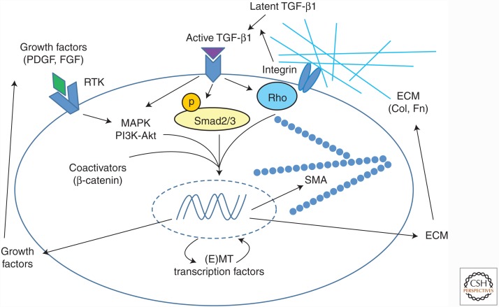
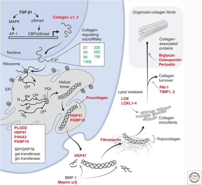
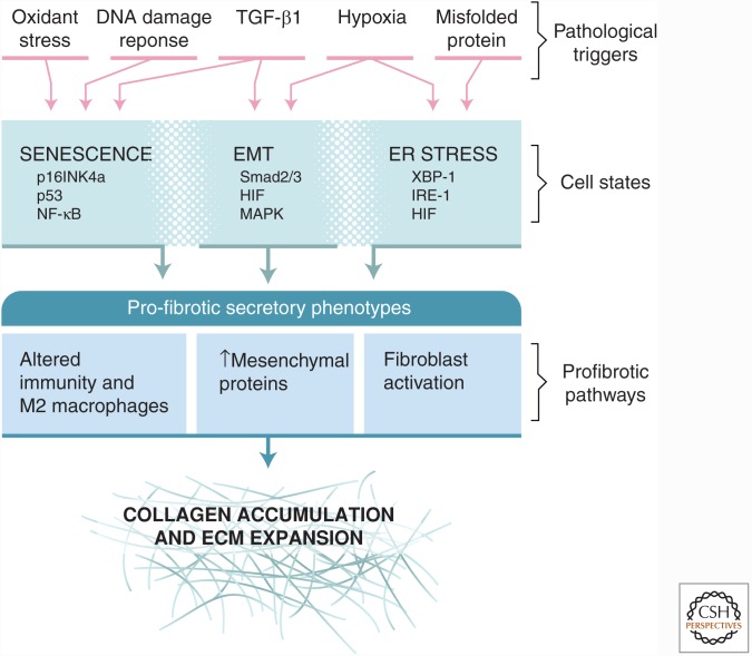
Activation of TGF-β1 initiates a program of temporary collagen accumulation important to wound repair in many organs. However, the outcome of temporary extracellular matrix strengthening all too frequently morphs into progressive fibrosis, contributing to morbidity and mortality worldwide. To avoid this maladaptive outcome, TGF-β1 signaling is regulated at numerous levels and intimately connected to feedback signals that limit accumulation. Here, we examine the current understanding of the core functions of TGF-β1 in promoting collagen accumulation, parallel pathways that promote physiological repair, and pathological triggers that tip the balance toward progressive fibrosis. Implicit in better understanding of these processes is the identification of therapeutic opportunities that will need to be further advanced to limit or reverse organ fibrosis.


Figures

Figure 1.
Figure 1.
Epithelial and mesenchymal cell fibrogenic activation by TGF-β1: costimulation by other signaling inputs and opportunities for feed-forward activation. Latent TGF-β is activated by αv integrins. Active TGF-β binds to its receptors leading to activation of canonical Smad signaling and Smad-independent signaling pathways, including MAPK pathways and small GTPases such as RhoA. Inputs from other signaling pathways converge on these pathways to regulate the TGF-β response. These are activated by other growth factors, such as PDGF, which generally signal through RTKs and through MAPK, integrin-mediated mechanotransduction mediated by Rho family GTPases, and coactivators such as β-catenin. These signaling factors lead to expression of profibrotic genes such as those encoding α-SMA, ECM proteins, and secreted cytokines and growth factors, which further modify the fibrogenic effector cell response. The expression of transcription factors involved in epithelial or mesenchymal cell transition into an activated state is also induced. PDGF, platelet-derived growth factor; FGF, fibroblast growth factor; RTK, receptor tyrosine kinase; MAPK, mitogen-activated protein kinase; (E)MT, (epithelial–) mesenchymal transition; Col, collagen; Fn, fibronectin; SMA, α-smooth muscle actin; ECM, extracellular matrix.
Figure 2.
Figure 2.
TGF-β1-induced collagen expression program. The schematic illustrates the proteins and microRNAs that are coordinately regulated by TGF-β1 to effect tissue collagen accumulation. Proteins whose expression is activated by TGF-β1 are highlighted in red, indicating that the expression of more than a dozen proteins is induced along with that of collagen and is required for substantial collagen expression and tissue accumulation. These proteins act at nearly every stage of collagen processing, from posttranslational proline and lysine hydroxylations by procollagen-lysine 2-oxogluterate 5-deoxygenase (PLOD2) and prolyl-4-hydroxylase (P4HA3), and glycosylation in the endoplasmic reticulum (ER), to required chaperones (HSP47 and FKBP10) to sustain the trimeric collagen structure and prevent premature fibril formation during passage through the secretory machinery. Additional, constitutively expressed proteins, such as protein disulfide isomerase (PDI), and quality control sensors, such as BiP/GRP78, are also important for collagen folding and subsequent expression. Once secreted, procollagen is proteolytically processed to generate tropocollagen that then spontaneously begins to self-associate into microfibrils. The final physical form and extent of stable, matrix fibrillar collagens depend heavily on additional TGF-β1-induced proteins such as fibronectin, the lysyl oxidases, and inhibitors of collagen turnover, plasminogen activator inhibitor 1 (PAI-1) and tissue inhibitor of metalloproteinases 1 and 3 (TIMP1, TIMP3). Finally, proteins such as biglycan and periostin associate with mature fibrils and control packing and organization. Collectively, this reprogramming of cells can be considered as the TGF-β1-induced collagen expression program.
Figure 3.
Figure 3.
Diverse pathways of epithelial responses to stress converge on profibrotic secretory phenotypes supporting extracellular matrix (ECM) expansion. The figure highlights three major epithelial states resulting from different pathological triggers: epithelial–mesenchymal transition (EMT), senescence, and endoplasmic reticulum (ER) stress with unfolded protein responses (UPRs). Each epithelial state has its characteristic transcriptional drivers. All of these states elicit secretory responses that share the potential to promote collagen accumulation. Distinctions among the secretory responses are still not completely defined and represent opportunities to better understand the connections between epithelial and mesenchymal biology in the context of cell stress. As discussed, TGF-β1 is a major driver of fibroblast activation and collagen secretion. Like epithelial cells, fibroblasts also respond to many of the same pathological triggers with activation of senescent or UPR signaling pathways, potentially further promoting fibrosis.

Similar articles

Cited by

References

    1. Acloque H, Adams MS, Fishwick K, Bronner-Fraser M, Nieto MA. 2009. Epithelial–mesenchymal transitions: The importance of changing cell state in development and disease. J Clin Invest 119: 1438–1449. - PMC - PubMed
    1. Adamali HI, Maher TM. 2012. Current and novel drug therapies for idiopathic pulmonary fibrosis. Drug Des Devel Ther 6: 261–272. - PMC - PubMed
    1. Ahluwalia N, Shea BS, Tager AM. 2014. New therapeutic targets in idiopathic pulmonary fibrosis. Aiming to rein in runaway wound-healing responses. Am J Respir Crit Care Med 190: 867–878. - PMC - PubMed
    1. Akagi K, Sandig V, Vooijs M, Van der Valk M, Giovannini M, Strauss M, Berns A. 1997. Cre-mediated somatic site-specific recombination in mice. Nucleic Acids Res 25: 1766–1773. - PMC - PubMed
    1. Al-Alawi M, Hassan T, Chotirmall SH. 2014. Transforming growth factor β and severe asthma: A perfect storm. Respir Med 108: 1409–1423. - PubMed

Publication types