. 2017 Aug;31(8):3622-3635.
doi: 10.1096/fj.201700092R. Epub 2017 Apr 21.

Folate-dependent methylation of septins governs ciliogenesis during neural tube closure

Affiliations

Folate-dependent methylation of septins governs ciliogenesis during neural tube closure

Manami Toriyama et al. FASEB J. 2017 Aug.

Abstract

Periconception maternal folic acid (vitamin B9) supplementation can reduce the prevalence of neural tube defects (NTDs), although just how folates benefit the developing embryo and promote closing of the neural tube and other morphologic processes during development remains unknown. Folate contributes to a 1-carbon metabolism, which is essential for purine biosynthesis and methionine recycling and affects methylation of DNA, histones, and nonhistone proteins. Herein, we used animal models and cultured mammalian cells to demonstrate that disruption of the methylation pathway mediated by folate compromises normal neural tube closure (NTC) and ciliogenesis. We demonstrate that the embryos with NTD failed to adequately methylate septin2, a key regulator of cilium structure and function. We report that methylation of septin2 affected its GTP binding activity and formation of the septin2-6-7 complex. We propose that folic acid promotes normal NTC in some embryos by regulating the methylation of septin2, which is critical for normal cilium formation during early embryonic development.-Toriyama, M., Toriyama, M., Wallingford, J. B., Finnell, R. H. Folate-dependent methylation of septins governs ciliogenesis during neural tube closure.

Keywords: epigenetics; folate transport; neural tube defect; septin2.


Figures

Figure 1.
Figure 1.
slc19a1 conditional-knockout mice show neural tube defects. A) The endogenous slc19a1 gene comprising exon 6 (top). After homologous recombination, the neomycin phosphotransferase (Neo) cassette was initially flanked by the FRT site and subsequently crossed with Cre line mice (bottom). B) PCR genotyping was performed using primer 1 (P1, A) and primer 2 (P2, A). Genomic DNA was extracted from the yolk sac or tail. C) Scheme of crossbreeding. Slc19a1 flox/flox mice were crossed with RFC1+/−;E2A-Cre mice. Embryos were isolated and observed at E9.5. D, E) RFC1 flox/;E2A-cre+/− embryo (E) exhibited an open neural tube (asterisk) compared with wild-type (D). F) Scheme of crossbreeding. Slc19a1 flox/flox mice were crossed with slc19a1 +/−;Wnt1-Cre+/− mice. Embryos were isolated and observed at E10.5, E13.5, E16.5, and E18.5. G) The comparison image between wild-type (right) and slc19a1 flox/;Wnt1-cre+/− (left) embryo. H) A magnified image of a slc19a1-knockout embryo; asterisk: open neural tube. I) Scheme of crossbreeding. Slc19a1 flox/flox mice were crossed with slc19a1+/−;Ttr-Cre+/− mice. Embryos were isolated and observed at E9.5, E13.5, and E18.5. J, K) The comparison image with wild-type (left) and slc19a1 flox/;Ttr1-cre+/− (right) embryo at E13.5; asterisk: open neural tube.
Figure 2.
Figure 2.
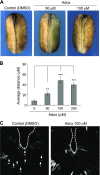
The methylation pathway is essential for neural tube closure in Xenopus. A) Dorsal view of stage 19 Xenopus embryos. Dashed lines: the medial side of each neural fold. Neural folds were closely opposed in the control embryo. After adox treatment, the neural tube remained open. B) The average distance between neural folds was 8.06 ± 2.63 mm (n = 25) in control embryos and 22.25 ± 4.27 (n = 33), 47.96 ± 6.74 (n = 29), and 39.5 ± 4.67 mm (n = 31) in 50, 100, and 200 µM adox-treated embryos, respectively. Mean values with bars representing sem are shown. **P < 0.01, ***P < 0.001 (Student’s t test). C) Primary cilia in Xenopus embryos. Arrows: cilia stained by acetylated tubulin.
Figure 3.
Figure 3.
Folate promotes ciliogenesis via methylation pathway. A) MEFs were grown in RPMI1640 folic-acid–free medium containing 200 µg/ml (top) or 0 µg/ml folinic acid, a 5-formyl derivative of tetrahydrofolic acid (bottom) for 2 d. Cells were cultured in serum-starved RPMI1640 medium containing 200 µg/ml (top) or 0 µg/ml folinic acid (bottom), for 16 h before fixing. Subsequently, the cells were immunostained with anti-acetylated tubulin (acetyl tub, ciliary marker; green) antibody. Nuclei were stained with DAPI (blue). B) Ciliated cells (A) were counted and graphed. Six different experiments were performed. C) MEFs were treated with DMSO as the control (top) or 10 µM adox, methylation inhibitor (bottom). Cilia were immunostained with anti-acetylated tubulin antibody (green). Nuclei were stained with DAPI (blue). D) The number of ciliated cells (C) was counted and graphed. Three different experiments were performed. E, F) Primary MEFs were isolated from E9.5 slc19a1 flox/flox embryos, then infected for 3 d with a retrovirus harboring GFP as control (E) or GFP with Cre recombinase (F). Cells were grown with RPMI1640, folic-acid–free medium containing 200 µg/ml folinic acid and 10% dialyzed FBS for 2 d. Sixteen hours before fixation, medium was changed to FBS-free RPMI1640 medium containing 200 µg/ml folinic acid. DMSO (top) or 40 µM adox (bottom) treatment was administered for 16 h before fixing, at which time the cells were immunostained with anti-arl13b (green) and DAPI (blue). G) Number of ciliated cells (E, F) were counted and graphed. *P < 0.05, **P < 0.01 (Student’s t test). Error bars ± se.
Figure 4.
Figure 4.
Folate regulates Shh signaling via methylation pathway. A) Western blot analysis for Gli3 (top band; full-length Gli3, bottom band; truncated Gli3) and β-tubulin in HEK293T cell lysate. HEK293T cells were grown in RPMI1640 medium containing 200 µg/ml folinic acid for 48 h. Adox (20 µM) was added 16 h before the cell lysate was prepared. DMSO was added as a control. B) Relative amount of truncated Gli3 (A) was calculated by normalizing to Gli3 full-length expression and then graphed. Three different experiments were performed. C) HEK293T cells were treated with 0–80 µM adox for 16 h before the cell lysate was prepared. Western blot analysis for Gli3 or β-tubulin was shown. D) Relative amount of truncated Gli3 (C) was calculated and graphed. Three different experiments were performed. E) Slc19a1-knockout mouse embryos were isolated at E9.5. After genotyping using PCR, Western blot analysis for Gli3 and β-tubulin was performed, as in A. *P < 0.05, **P < 0.01, ***P < 0.001 (Student’s t test). Error bars ± se.
Figure 5.
Figure 5.
Human septin2 is methylated via folate pathway. A) Western blot analysis for methylated lysine (meK) and FLAG. HEK293T cells were transfected with FLAG-septin2. Thirty-six hours after transfection, the cells were treated with 40 µM adox for 16 h. After preparation of cell lysates, FLAG-septin2 was immunoprecipitated using FLAG antibody. Mouse IgG antibody was used as the control for the immunoprecipitation. B) HEK293T cells expressing FLAG-septin2 were grown in RPMI1640 medium containing either 200 µg/ml folinic acid or folate depletion medium for 2 d. After immunoprecipitation of FLAG-septin2 from cell lysate, Western blot analysis was performed to detect meK and FLAG. C, D) Spectrum data for septin2 peptides containing Lys 183 (C) and Arg 300 (D) by LC-MS/MS. FLAG-septin2 purified from HEK293T cells were digested by trypsin, and mass spectrometry was performed. Molecular weight increase secondary to methylation (MW 14) at both residues was identified. E, F) Sequence alignment of septin2 from human to yeast. Methylated lysine (E) and dimethylated arginine (F) are highlighted in yellow. Highly conserved residues from human to yeast are highlighted in blue. Nonconcordance with human residues is highlighted in green.
Figure 6.
Figure 6.
Methylation of septin2 regulates GTP binding affinity and formation of septin complex. A) HEK293T cells were transfected with FLAG-septin2 and cultured in RPMI1640 medium containing 200 µg/ml folinic acid for 2 d. The cells were treated with 40 µM adox for 16 h before they were collected. After immunoprecipitation of FLAG-septin2, GTP binding assays were performed. The biotinylated GTP binding form of FLAG-septin2 was specifically precipitated using streptavidin beads. Western blot analysis of FLAG-septin2 in precipitant or 5% input was shown. B) Relative amount of GTP binding form of septin2 (A) was calculated and graphed. Band intensity was normalized to input. C) HEK293T cells were transfected with FLAG-septin2 mutants, and septin2 proteins were purified by immunoprecipitation. GTP binding assays were performed, and GTP binding septin2 was detected by FLAG antibody. D) Relative amount of GTP binding form of septin2 shown in C. Band intensity was normalized to input. Three different experiments were performed. E) HEK293T cells were cotransfected with FLAG-septin2 and GFP-septin6. Sixteen hours before cell lysates were prepared, 40 µM adox was added to the medium. FLAG-septin2 was continuously immunoprecipitated by FLAG antibody, and coprecipitated proteins were detected by Western blot. F) Binding affinity of GFP-septin6 and endogenous septin7 with FLAG-septin2 (E) was graphed. G) FLAG-septin2 mutants in HEK293T cells were immunoprecipitated with FLAG antibody and coprecipitated GFP-septin6 or endogenous septin7 was detected. H) Binding affinity of GFP-septin6 and endogenous septin7 with FLAG-septin2 (G) was graphed. *P < 0.05, **P < 0.01, ***P < 0.001 (Student’s t test). Error bars ± se.
Figure 7.
Figure 7.
Methylation at Lys 183 of septin2 prevents aggregation thereby promoting ciliogenesis. A) Cell localization of GFP or GFP-septin2. NIH3T3 cells were transfected with GFP or GFP-septin2. Two days after transfection, cells were cultured with serum-starved medium containing 20 µM adox for 16 h. DMSO was used as the control. Primary cilia were detected with anti-acetylated tubulin (acetyl tub, red) antibody. Nuclei were detected with DAPI (blue). Arrows: primary cilia; arrowheads: aggregated septin2. B) Number of aggregated GFP-septin2 shown in A were counted and graphed. C) Cell localization of GFP-septin2 mutants. Primary cilia and nuclei were detected with anti-acetylated tubulin antibody (red) or DAPI (blue), respectively. Arrows: primary cilia; arrowheads: aggregated septin2. D) Aggregated septin2 (C) was counted and its aggregation rate was calculated. Three different experiments were performed. E) Number of ciliated cells (C) were counted and graphed. Three different experiments were performed. F) HEK293T cells were transfected with FLAG-septin2. Gli3, FLAG, and β-tubulin in cell lysate were detected by Western blot. G) Relative amount of truncated Gli3 (F) was calculated by normalizing to Gli3 full-length and graphed. *P < 0.05, **P < 0.01 (Student’s t test). Error bars ± se.
Figure 8.
Figure 8.
Folate-methylation pathway for ciliogenesis during embryogenesis. Folate signaling contributes to methylation of septin2, which regulates formation of the septin2-6-7 complex and ciliogenesis.

Similar articles

Cited by

References

    1. Cearns M. D., Escuin S., Alexandre P., Greene N. D., Copp A. J. (2016) Microtubules, polarity and vertebrate neural tube morphogenesis. J. Anat. 229, 63–74 - PMC - PubMed
    1. Golden J. A., Chernoff G. F. (1993) Intermittent pattern of neural tube closure in two strains of mice. Teratology 47, 73–80 - PubMed
    1. Karfunkel P. (1974) The mechanisms of neural tube formation. Int. Rev. Cytol. 38, 245–271 - PubMed
    1. Copp A. J., Greene N. D., Murdoch J. N. (2003) The genetic basis of mammalian neurulation. Nat. Rev. Genet. 4, 784–793 - PubMed
    1. Greene N. D., Copp A. J. (2014) Neural tube defects. Annu. Rev. Neurosci. 37, 221–242 - PMC - PubMed

Publication types

MeSH terms

LinkOut - more resources