

Review
doi: 10.1161/STROKEAHA.116.012784.
Intracranial Dural Arteriovenous Fistulae
Affiliations
- PMID: 28432263
- PMCID: PMC5435465
- DOI: 10.1161/STROKEAHA.116.012784
Item in Clipboard
Review
Intracranial Dural Arteriovenous Fistulae
Stroke.
2017 May.
No abstract available
Keywords: Borden–Shucart; Cognard; Zipfel, Barrow; cortical venous drainage; dural arteriovenous fistula; intracranial; shunt.
Figures
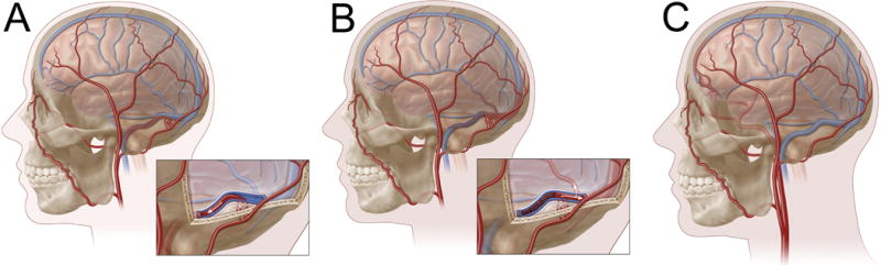
Borden-Shucart classification of intracranial dAVF. In Type I fistulae (A), progressive venous sinus stenosis and/or occlusion results in venous congestion and anastomoses between meningeal arteries (e.g., trans-osseous feeders from the occipital artery, as depicted here) and a dural venous sinus (e.g., transverse-sigmoid junction, as depicted here). Venous flow is entirely anterograde and cortical venous drainage (CVD) is absent. In Type II fistulae (B), progressively worsening venous hypertension results in both anterograde (into sinus) and retrograde (CVD) venous outflow. In Type III lesions (C), a meningeal artery shunts directly into a cortical vein (with CVD). Venous outflow, by definition, is entirely retrograde. In this example, ethmoidal branches from the ophthalmic artery shunt directly into a cortical frontal vein resulting in a venous varix.
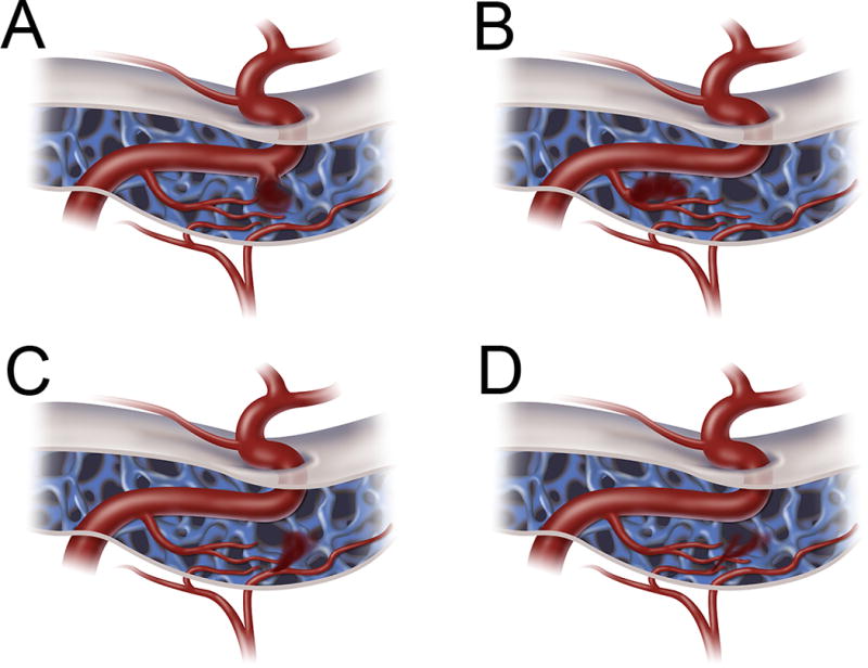
Barrow classification of CCFs. Type A (A) CCFs are direct fistulas between the intracavernous ICA and the CS (most common). Type B (B) CCFs are indirect fistulas between meningeal branches of the intracavernous ICA and the CS. Type C (C) CCFs are indirect fistulas between meningeal branches of the ECA and the CS. Type D (D) CCFs are indirect fistulas between both meningeal branches of the intracavernous ICA and meningeal braches of the ECA and the CS.
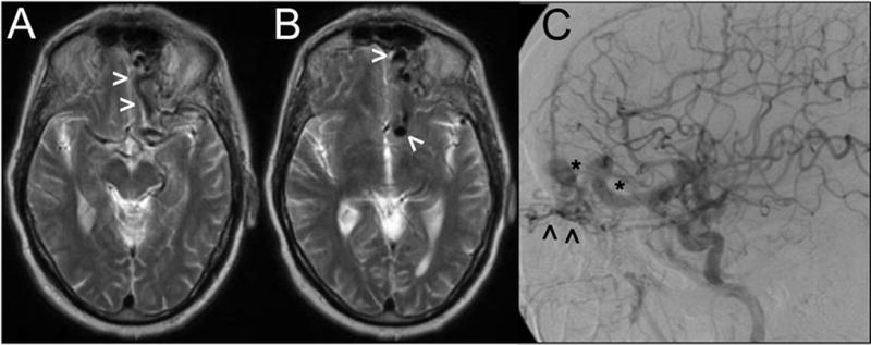
Axial sections from a T2-weighted MRI study (A, B) showing large vascular flow voids (arrowheads) near the left anterior fossa floor. Digital subtraction angiography (lateral view following a left CCA contrast injection) demonstrates a Borden-Shucart Type III dAVF fed by ethmoidal branches from the ophthalmic artery (arrowheads) and draining into enlarged, arterialized cortical frontal veins with an associated venous ectasia and varices (asterisks).
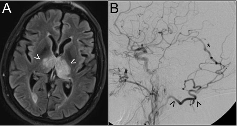
This patient presented with rapidly-progressive dementia. Axial MR imaging with FLAIR sequences (A) demonstrates bi-thalamic edema (arrowheads) as a result of venous hypertension. Digital subtraction angiography (B; lateral view following a left CCA contrast injection) shows a high-grade dAVF fed by the occipital artery (arrowhead) with early arteriovenous shunting into the deep venous system (e.g., vein of Galen, asterisk). Endovascular embolization resulted in complete fistula obliteration and resolution of clinical and radiographic abnormalities.
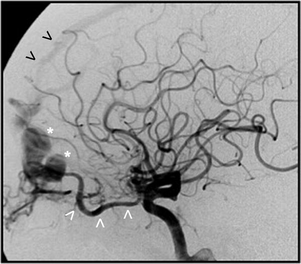
Digital subtraction angiogram (lateral view after a left CCA contrast injection) shows a Borden-Shucart Type III dAVF supplied via ethmoidal branches from a hypertrophic ophthalmic artery (white arrowheads) and draining into a cortical frontal vein causing venous ectasia and a large venous varix (asterisk). Early filling of the anterior third of the superior sagittal sinus can also be appreciated (black arrowheads).
References
-
- Newton TH, Cronqvist S. Involvement of dural arteries in intracranial arteriovenous malformations 1. Radiology. 1969;93:1071–1078. - PubMed
-
- Brown RD, Jr, Wiebers DO, Nichols DA. Intracranial dural arteriovenous fistulae: Angiographic predictors of intracranial hemorrhage and clinical outcome in nonsurgical patients. Journal of neurosurgery. 1994;81:531–538. - PubMed
-
- Söderman M, Pavic L, Edner G, Holmin S, Andersson T. Natural history of dural arteriovenous shunts. Stroke. 2008;39:1735–1739. - PubMed
-
- Strom RG, Botros JA, Refai D, Moran CJ, Cross DT, III, Chicoine MR, et al. Cranial dural arteriovenous fistulae: Asymptomatic cortical venous drainage portends less aggressive clinical course. Neurosurgery. 2009;64:241–248. - PubMed
Publication types
MeSH terms
Grants and funding
LinkOut - more resources
Full Text Sources
Other Literature Sources
