

Establishment of an in vitro culture model of theca cells from hierarchical follicles in ducks
- PMID: 28432272
- PMCID: PMC5426285
- DOI: 10.1042/BSR20160491
Establishment of an in vitro culture model of theca cells from hierarchical follicles in ducks
Abstract
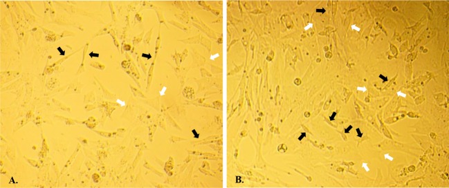
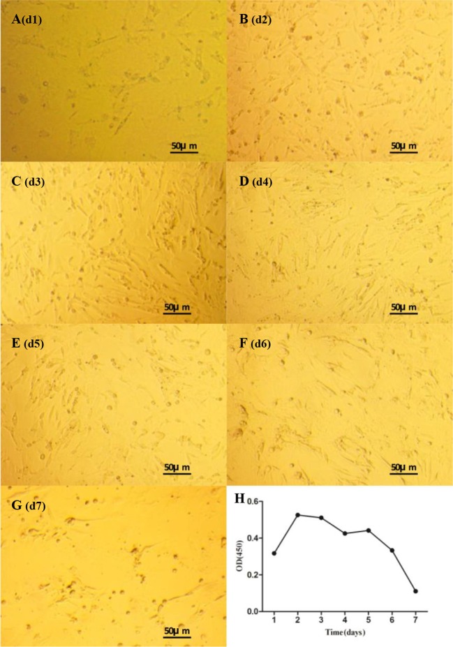
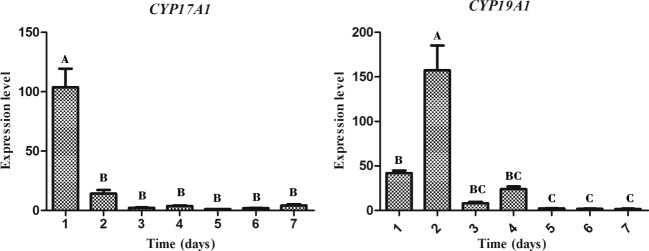
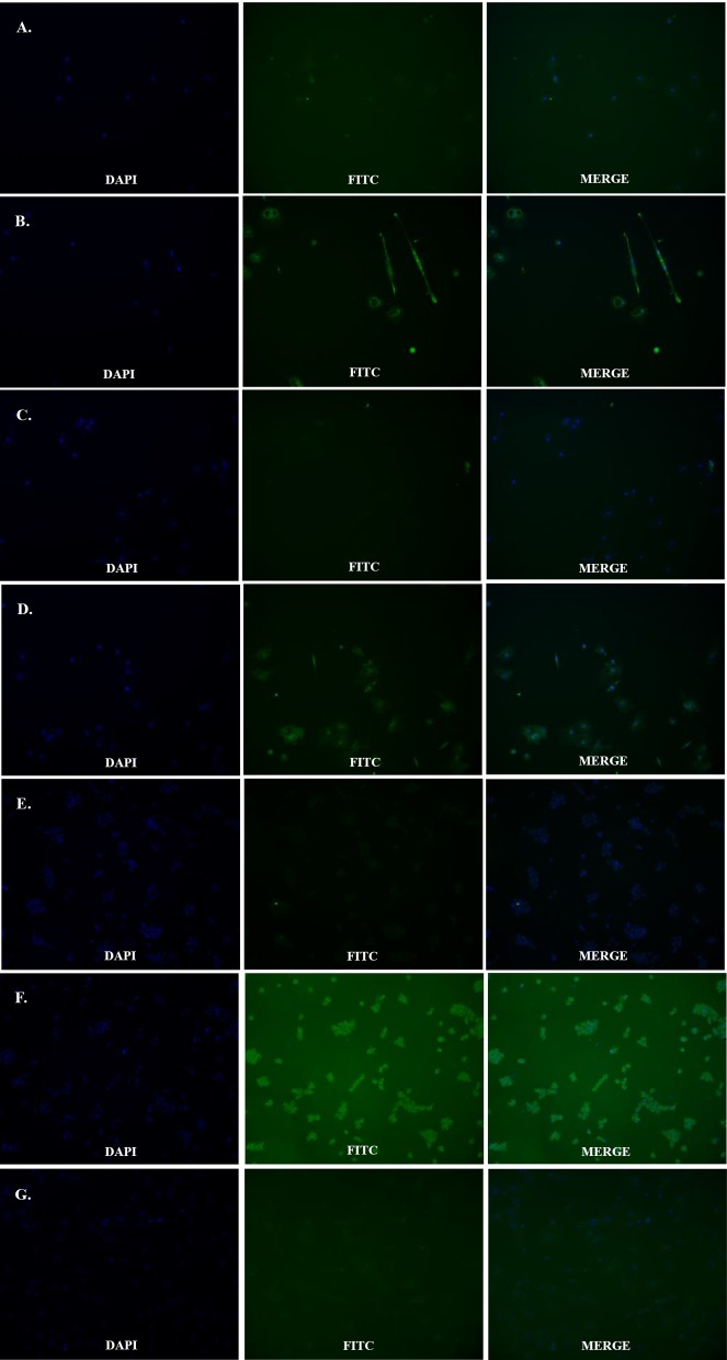
Theca cells, including theca interna cells and theca externa cells, are vital components of ovarian follicles. The aim of the present study is to identify a reliable method for the in vitro culture of theca cells from duck ovarian hierarchical (F4-F2) follicles. We improved the method for cell separation by using trypsin to further remove granular cells, and we increased the concentration of fetal bovine serum used in in vitro culture to improve cytoactivity. Cell antibody immunofluorescence (IF) showed that all inoculated cells could be stained by the CYP17A1/19A1 antibody but not by the FSHR antibody, which could stain granulosa cells. Furthermore, morphological differences were observed between the outlines of theca interna and externa cells and in their nuclei. Growth curve and CYP17A1/19A1 mRNA relative expression analyses suggested that the growth profile of theca interna cells may have been significantly different from that of theca externa cells in vitro Theca interna cells experienced the logarithmic phase on d1-d2, the plateau phase on d2-d3, and the senescence phase after d3, while theca externa cells experienced the logarithmic phase on d1-d3, the plateau phase on d3-d5, and the senescence phase after d5. Taken together, these results suggested that we have successfully established a reliable theca cell culture model and further defined theca cell characteristics in vitro.
Keywords: characteristics; culture model; duck; theca cell.
© 2017 The Author(s).
Conflict of interest statement
The authors declare that there are no competing interests associated with the manuscript.
Figures
Similar articles
-
[Expression of mRNAs for GHR, IGF-IR, FSHR and LHR in granulosa and theca layers of ovarian follicles of Shaoxing ducks].Yi Chuan Xue Bao. 2003 Sep;30(9):840-6. Yi Chuan Xue Bao. 2003. PMID: 14577376 Chinese.
-
Growth differentiation factor 9 (GDF9) stimulates proliferation and inhibits steroidogenesis by bovine theca cells: influence of follicle size on responses to GDF9.Biol Reprod. 2008 Feb;78(2):243-53. doi: 10.1095/biolreprod.107.063446. Epub 2007 Oct 24. Biol Reprod. 2008. PMID: 17959852
-
Steroidogenesis by equine preovulatory follicles: relative roles of theca interna and granulosa cells.Endocrinology. 1991 Feb;128(2):1159-66. doi: 10.1210/endo-128-2-1159. Endocrinology. 1991. PMID: 1989851
-
Amount of mRNA and localization of vascular endothelial growth factor and its receptors in the ovarian follicle during estrous cycle of water buffalo (Bubalus bubalis).Anim Reprod Sci. 2013 Mar;137(3-4):163-76. doi: 10.1016/j.anireprosci.2013.01.004. Epub 2013 Jan 17. Anim Reprod Sci. 2013. PMID: 23375984
-
Microvascular distribution and vascular endothelial growth factor expression in bovine cystic follicles.Domest Anim Endocrinol. 2005 Nov;29(4):634-45. doi: 10.1016/j.domaniend.2005.04.007. Epub 2005 May 31. Domest Anim Endocrinol. 2005. PMID: 15950432
Cited by
-
Proteome of granulosa cells lipid droplets reveals mechanisms regulating lipid metabolism at hierarchical and pre-hierarchical follicle in goose.Front Vet Sci. 2025 Mar 31;12:1544718. doi: 10.3389/fvets.2025.1544718. eCollection 2025. Front Vet Sci. 2025. PMID: 40230795 Free PMC article.
-
Transcriptome Analysis of circRNA and mRNA in Theca Cells during Follicular Development in Chickens.Genes (Basel). 2020 Apr 29;11(5):489. doi: 10.3390/genes11050489. Genes (Basel). 2020. PMID: 32365656 Free PMC article.
-
Comparative Analysis Among Different Species Reveals That the Androgen Receptor Regulates Chicken Follicle Selection Through Species-Specific Genes Related to Follicle Development.Front Genet. 2022 Jan 3;12:752976. doi: 10.3389/fgene.2021.752976. eCollection 2021. Front Genet. 2022. PMID: 35046998 Free PMC article.
-
Combined analyses of mRNA and miRNA transcriptome reveal the molecular mechanisms of theca cells physiological differences in geese follicular selection stage.Poult Sci. 2024 Dec;103(12):104402. doi: 10.1016/j.psj.2024.104402. Epub 2024 Oct 10. Poult Sci. 2024. PMID: 39510003 Free PMC article.
-
Characterization of the In Vitro Cultured Ovarian Cells in the Asian Yellow Pond Turtle (Mauremys mutica).Biology (Basel). 2022 Sep 26;11(10):1404. doi: 10.3390/biology11101404. Biology (Basel). 2022. PMID: 36290308 Free PMC article.
References
-
- Nitta H., Osawa Y. and Bahr J.M. (1991) Immunolocalization of steroidogenic cells in small follicles of the chicken ovary: anatomical arrangement and location of steroidogenic cells change during follicular development. Domest. Anim. Endocrinol. 8, 587–594 - PubMed
-
- Lee K.A., Volentine K.K. and Bahr J.M. (1998) Two steroidogenic pathways present in the chicken ovary: theca layer prefers delta 5 pathway and granulosa layer prefers delta 4 pathway. Domest. Anim. Endocrinol. 15, 1–8 - PubMed
-
- Miyoshi T., Otsuka F., Inagaki K., Otani H., Takeda M., Suzuki J. et al. (2007) Differential regulation of steroidogenesis by bone morphogenetic proteins in granulosa cells: involvement of extracellularly regulated kinase signaling and oocyte actions in follicle-stimulating hormone-induced estrogen production. Endocrinology 148, 337–345 - PubMed
-
- Otsuka F. (2010) Multiple endocrine regulation by bone morphogenetic protein system. Endocr. J. 57, 3–14 - PubMed
MeSH terms
Substances
LinkOut - more resources
Full Text Sources
Other Literature Sources
Medical
Miscellaneous