. 2017 Apr 21;7(1):1037.
doi: 10.1038/s41598-017-00902-5.

Growth Differentiation Factor-15 Deficiency Augments Inflammatory Response and Exacerbates Septic Heart and Renal Injury Induced by Lipopolysaccharide

Affiliations

Growth Differentiation Factor-15 Deficiency Augments Inflammatory Response and Exacerbates Septic Heart and Renal Injury Induced by Lipopolysaccharide

Palida Abulizi et al. Sci Rep. .

Abstract

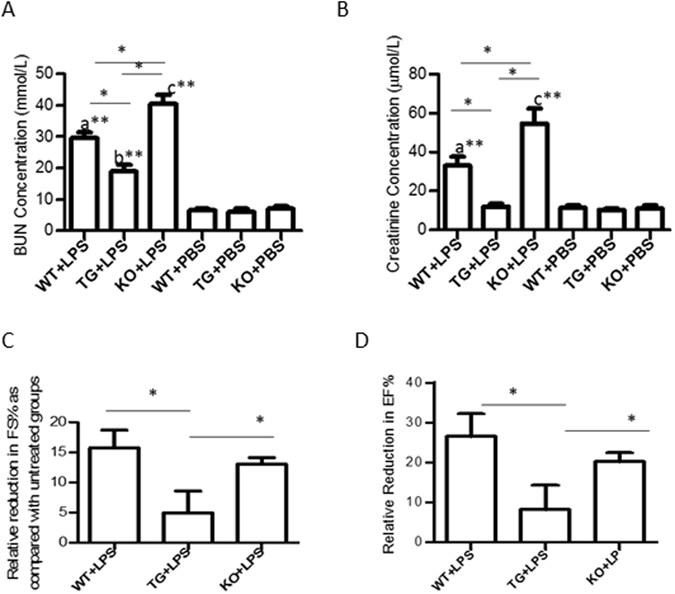
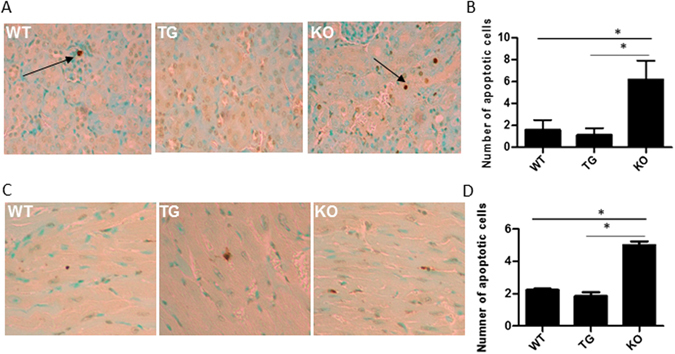
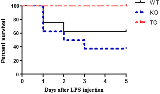
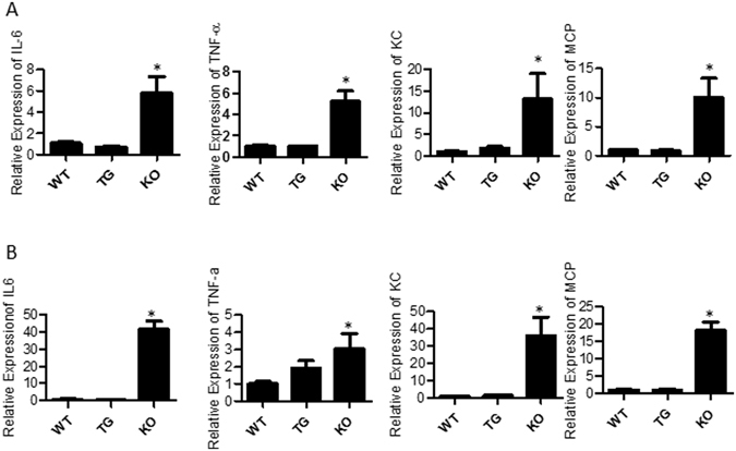
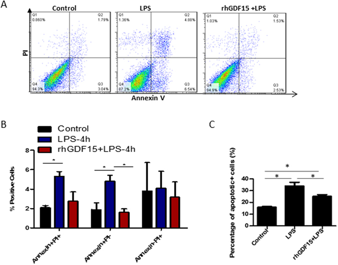
Septic acute kidney injury (AKI) and myocardial dysfunction are leading causes of mortality with no accepted method of therapy. In this study we demonstrate the role of growth differentiating factor 15 (GDF15) in septic AKI and myocardial dysfunction using a murine lipopolysaccharide (LPS)-induced sepsis model and an in vitro cell culture system. Data show that GDF15 deficiency augments inflammatory response and exacerbates renal and cardiac injury induced by LPS, while over-expression of GDF15 protects the kidney and heart from LPS-induced organ dysfunction. Therefore, this study highlights the therapeutic potential of GDF15 in the treatment of endotoxin-induced sepsis.


Conflict of interest statement

The authors declare that they have no competing interests.

Figures

Figure 1
Figure 1
GDF15 deficiency exacerbates cardiac and renal injury after LPS administration. WT, GDF15 TG and GDF15 KO mice were i.p. injected with 4 mg/kg of LPS (WT + LPS, TG + LPS and KO + LPS group) or PBS (WT, TG and KO group) for 16 hours. Blood was collected to measure concentrations of BUN (A) and serum creatinine (B) to assess renal function Ultrasound scanning was performed to measure EF% (C) and FS% (D) for heart function. After ultrasound scanning, mice were sacrificed. n = 9–10 for mice treated with LPS and n = 4/group for PBS injected groups in Fig. 1(A,B); n = 5 for (C,D); *P ≤ 0.05, **P ≤ 0.01. a: WT + LPS vs. either WT, TG or KO group; b: TG + LPS vs. either WT, TG or KO group; c: KO + LPS vs. either WT, TG or KO group.
Figure 2
Figure 2
GDF15 KO mice show greater levels of tubular damage and cardiac injury, and increased neutrophil infiltration. Sixteen hours after LPS treatment, kidneys and hearts were harvested from mice. Tissues from all groups were sectioned and stained with H&E and detected MPO activity. Images were taken from kidney cortex at 200 X magnification. (A) Representative HE staining images of kidney tissues. (B) Injury score for kidney tissues. (C) Representative MPO images of kidney tissues. (D) MPO score for kidney tissues. (E) Representative HE staining images of heart tissues. (F) Injury score for heart tissues. (G) Representative MPO images of heart tissues. (H) MPO score for heart tissues. n = 6 for HE staining. n = 4 for MPO assays. *P ≤ 0.05.
Figure 3
Figure 3
GDF15 KO mice have increased levels of apoptotic cells in kidney (A,B) and heart tissues (C,D). Formalin fixed, paraffin embedded sections of GDF15 WT, TG and KO mice treated with LPS were stained for the TUNEL assay. The number of stained cells were counted for each slide across ten sections at 200 X magnification and averaged. (A,C) are representative images. (B,D) are summarized data. n = 4;*P ≤ 0.05, **P ≤ 0.01, ***P ≤ 0.001.
Figure 4
Figure 4
Over expression of GDF15 prevents LPS-induced mortality of animals. WT, TG and KO mice (n = 8) were i.p. injected with 20 mg/kg LPS. Animal health and survival was observed every 12 h in for 5 days after LPS injection.
Figure 5
Figure 5
Inflammatory gene expression in kidney and heart tissue. WT, TG and KO mice were i.p injected with LPS. RNA was extracted from the cortex of the kidney and heart 16 h after LPS treatment. qRT-PCR was performed using SYBR green to detect the expression of IL-6, TNF-α, KC and MCP-1 and the housekeeping gene GAPDH was used as the internal control. Data is expressed using the ∆∆CT method and normalized to WT + LPS. (A) Kidney tissues (B) Heart tissues. n = 3–5 = ;*P ≤ 0.05.
Figure 6
Figure 6
Prior treatment with rhGDF15 reduces the amount of early apoptotic cells seen 4 hours after LPS treatment. Primary kidney tubular cells and primary cardiomyocytes from WT mice were cultured and treated with 25 µg/ml LPS alone for 4 hs, rhGDF15 two hours prior to LPS treatment or were untreated. Cells were then collected, stained with Annexin V and PI and detected using flow cytometry. Representative dot plots are shown for each treatment group (A). Summarized data for kidney cells are shown in (B) and for heart cells in (C). Data are representative of 3 independent experiments; *P ≤ 0.05.

References

    1. Bhan, C., Dipankar, P., Chakraborty, P. & Sarangi, P.P. Role of cellular events in the pathophysiology of sepsis. Inflammation research: official journal of the European Histamine Research Society … [et al.] (2016). - PubMed
    1. Husak, L. et al. National analysis of sepsis hospitalizations and factors contributing to sepsis in-hospital mortality in Canada. Healthcare quarterly (Toronto, Ont.) 13 Spec No, 35–41 (2010). - PubMed
    1. Chang HJ, Lynm C, Glass RM. JAMA patient page. Sepsis. Jama. 2010;304:1856. doi: 10.1001/jama.304.16.1856. - DOI - PubMed
    1. Zarbock A, Gomez H, Kellum JA. Sepsis-induced acute kidney injury revisited: pathophysiology, prevention and future therapies. Current opinion in critical care. 2014;20:588–595. doi: 10.1097/MCC.0000000000000153. - DOI - PMC - PubMed
    1. Zarjou A, Agarwal A. Sepsis and acute kidney injury. J Am Soc Nephrol. 2011;22:999–1006. doi: 10.1681/ASN.2010050484. - DOI - PubMed

Publication types

Substances

Grants and funding