. 2017 Jul:170:82-92.
doi: 10.1016/j.bandl.2017.03.012. Epub 2017 Apr 20.

Multimodal imaging of language reorganization in patients with left temporal lobe epilepsy

Affiliations

Multimodal imaging of language reorganization in patients with left temporal lobe epilepsy

Yu-Hsuan A Chang et al. Brain Lang. 2017 Jul.

Abstract

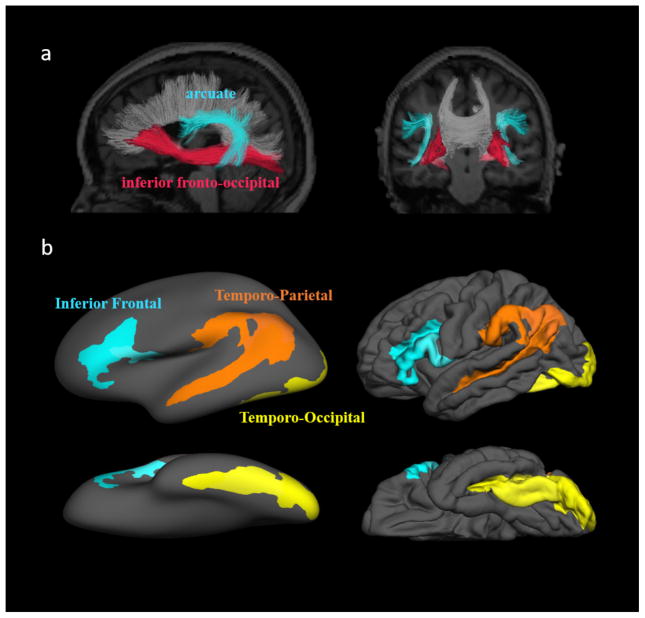
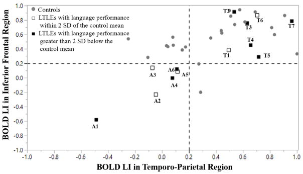
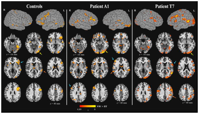
This study explored the relationships among multimodal imaging, clinical features, and language impairment in patients with left temporal lobe epilepsy (LTLE). Fourteen patients with LTLE and 26 controls underwent structural MRI, functional MRI, diffusion tensor imaging, and neuropsychological language tasks. Laterality indices were calculated for each imaging modality and a principal component (PC) was derived from language measures. Correlations were performed among imaging measures, as well as to the language PC. In controls, better language performance was associated with stronger left-lateralized temporo-parietal and temporo-occipital activations. In LTLE, better language performance was associated with stronger right-lateralized inferior frontal, temporo-parietal, and temporo-occipital activations. These right-lateralized activations in LTLE were associated with right-lateralized arcuate fasciculus fractional anisotropy. These data suggest that interhemispheric language reorganization in LTLE is associated with alterations to perisylvian white matter. These concurrent structural and functional shifts from left to right may help to mitigate language impairment in LTLE.

Keywords: Cortical thickness; DTI; Functional MRI; Language; Temporal lobe epilepsy.


Figures

Figure 1
Figure 1
a. The two white matter tracts of interest shown for a single individual. b. The three language regions of interest selected for the functional and volumetric MRI analyses. The inferior frontal region consists of the opercular, orbital, and triangular part of the inferior frontal gyrus (index 12, 13, and 14) and the inferior frontal sulcus (index 52) in the Destrieux altas. The temporo-parietal region consists of the angular gyrus (index 25), supramarginal gyrus (index 26), and superior temporal sulcus (index 73). The temporo-occipital region consists of the inferior occipital gyrus and sulcus (index 2), lateral occipito-temporal gyrus (index 21), occipital pole (index 42), anterior and posterior transverse collateral sulcus (index 50 and 51), and lateral occipito-temporal sulcus (index 60).
Figure 2
Figure 2
The plot for atypical (A) and typical (T) language dominance in both controls and patients with LTLE. The lower left quadrant indicates atypical language dominance. The empty square indicates that patient’s language performance was greater than 2 standard deviations below of the mean in the control group, whereas the solid square indicates that patient’ language performance was less than 2 standard deviations below the mean of the control.
Figure 3
Figure 3
The fMRI activation maps for the contrast of NW > FF in the control group, Patient A1, and Patient T7. The activation in the inferior frontal, temporo-parietal, and temporo-occipital regions were indicated by the cyan, orange, yellow arrows, respectively. The z coordinate was presented in Talairach space. p < .01, cluster size > 25 voxels, corrected α < .05.

References

    1. Adcock JE, Wise RG, Oxbury JM, Oxbury SM, Matthews PM. Quantitative fMRI assessment of the differences in lateralization of language-related brain activation in patients with temporal lobe epilepsy. Neuroimage. 2003;18(2):423–438. doi: 10.1016/s1053-8119(02)00013-7. - DOI - PubMed
    1. Ahmadi ME, Hagler DJ, McDonald CR, Tecoma ES, Iragui VJ, Dale AM, Halgren E. Side matters: Diffusion tensor imaging tractography in left and right temporal lobe epilepsy. American Journal of Neuroradiology. 2009;30(9):1740–1747. doi: 10.3174/ajnr.a1650. - DOI - PMC - PubMed
    1. Almairac F, Herbet G, Moritz-Gasser S, de Champfleur NM, Duffau H. The left inferior fronto-occipital fasciculus subserves language semantics: A multilevel lesion study. Brain Structure and Function. 2015;220(4):1983–1995. doi: 10.1007/s00429-014-0773-1. - DOI - PubMed
    1. Barnett A. Unpublished master’s thesis. University of Toronto; Toronto, Ontario, Canada: 2012. Memory functioning in patients with unilateral temporal lobe epilepsy: Neuroimaging indicators of functional integrity in the hippocampus and beyond.
    1. Bell B, Hermann B, Seidenberg M, Davies K, Cariski D, Rosenbek J, Woodard A, Rutecki P, Bishop M. Ipsilateral reorganization of language in early-onset left temporal lobe epilepsy. Epilepsy & Behavior. 2002;3(2):158–164. doi: 10.1006/ebeh.2002.0322. - DOI - PubMed

MeSH terms