Risk of Ocular Hypertension in Adults with Noninfectious Uveitis
- PMID: 28433444
- PMCID: PMC5522760
- DOI: 10.1016/j.ophtha.2017.03.041
Risk of Ocular Hypertension in Adults with Noninfectious Uveitis
Abstract
Purpose: To describe the risk and risk factors for ocular hypertension (OHT) in adults with noninfectious uveitis.
Design: Retrospective, multicenter, cohort study.
Participants: Patients aged ≥18 years with noninfectious uveitis seen between 1979 and 2007 at 5 tertiary uveitis clinics.
Methods: Demographic, ocular, and treatment data were extracted from medical records of uveitis cases.
Main outcome measures: Prevalent and incident OHT with intraocular pressures (IOPs) of ≥21 mmHg, ≥30 mmHg, and increase of ≥10 mmHg from documented IOP recordings (or use of treatment for OHT).
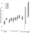
Results: Among 5270 uveitic eyes of 3308 patients followed for OHT, the mean annual incidence rates for OHT ≥21 mmHg and OHT ≥30 mmHg are 14.4% (95% confidence interval [CI], 13.4-15.5) and 5.1% (95% CI, 4.7-5.6) per year, respectively. Statistically significant risk factors for incident OHT ≥30 mmHg included systemic hypertension (adjusted hazard ratio [aHR], 1.29); worse presenting visual acuity (≤20/200 vs. ≥20/40, aHR, 1.47); pars plana vitrectomy (aHR, 1.87); history of OHT in the other eye: IOP ≥21 mmHg (aHR, 2.68), ≥30 mmHg (aHR, 4.86) and prior/current use of IOP-lowering drops or surgery in the other eye (aHR, 4.17); anterior chamber cells: 1+ (aHR, 1.43) and ≥2+ (aHR, 1.59) vs. none; epiretinal membrane (aHR, 1.25); peripheral anterior synechiae (aHR, 1.81); current use of prednisone >7.5 mg/day (aHR, 1.86); periocular corticosteroids in the last 3 months (aHR, 2.23); current topical corticosteroid use [≥8×/day vs. none] (aHR, 2.58); and prior use of fluocinolone acetonide implants (aHR, 9.75). Bilateral uveitis (aHR, 0.69) and previous hypotony (aHR, 0.43) were associated with statistically significantly lower risk of OHT.
Conclusions: Ocular hypertension is sufficiently common in eyes treated for uveitis that surveillance for OHT is essential at all visits for all cases. Patients with 1 or more of the several risk factors identified are at particularly high risk and must be carefully managed. Modifiable risk factors, such as use of corticosteroids, suggest opportunities to reduce OHT risk within the constraints of the overriding need to control the primary ocular inflammatory disease.
Copyright © 2017 American Academy of Ophthalmology. All rights reserved.
Conflict of interest statement
Figures


Comment in
-
Uveitis.Ophthalmologe. 2018 Sep;115(9):708-709. doi: 10.1007/s00347-018-0761-6. Ophthalmologe. 2018. PMID: 30187253 German. No abstract available.
References
-
- Kulkarni A, Barton K. Uveitic Glaucoma. In: Shaarawy T, editor. Glaucoma. 2. Vol. 1. London: Elsevier; 2015. pp. 410–24.
-
- Gordon MO, Beiser JA, Brandt JD, et al. The Ocular Hypertension Treatment Study: baseline factors that predict the onset of primary open-angle glaucoma. Arch Ophthalmol. 2002;120:714–20. - PubMed
-
- Miglior S, Pfeiffer N, Torri V, et al. Predictive factors for open-angle glaucoma among patients with ocular hypertension in the European Glaucoma Prevention Study. Ophthalmology. 2007;114:3–9. - PubMed
Publication types
MeSH terms
Substances
Grants and funding
LinkOut - more resources
Full Text Sources
Other Literature Sources
Miscellaneous

