. 2017 Jun;198(3):203-209.
doi: 10.1016/j.jsb.2017.04.006. Epub 2017 Apr 19.

Clostridium difficile toxin glucosyltransferase domains in complex with a non-hydrolyzable UDP-glucose analogue

Affiliations

Clostridium difficile toxin glucosyltransferase domains in complex with a non-hydrolyzable UDP-glucose analogue

Joseph W Alvin et al. J Struct Biol. 2017 Jun.

Abstract

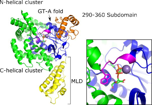
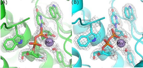
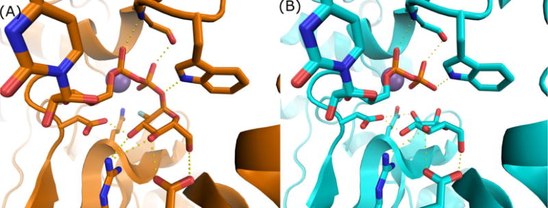
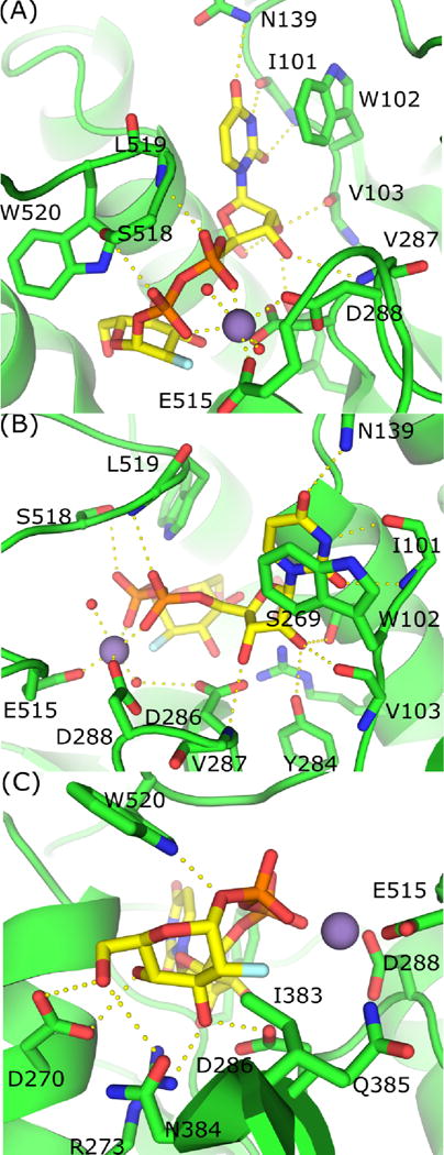
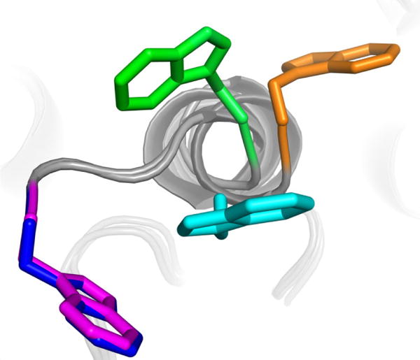
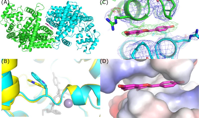
Clostridium difficile is the leading cause of hospital-acquired diarrhea and pseudomembranous colitis worldwide. The organism produces two homologous toxins, TcdA and TcdB, which enter and disrupt host cell function by glucosylating and thereby inactivating key signalling molecules within the host. As a toxin-mediated disease, there has been a significant interest in identifying small molecule inhibitors of the toxins' glucosyltransferase activities. This study was initiated as part of an effort to identify the mode of inhibition for a small molecule inhibitor of glucosyltransferase activity called apigenin. In the course of trying to get co-crystals with this inhibitor, we determined five different structures of the TcdA and TcdB glucosyltransferase domains and made use of a non-hydrolyzable UDP-glucose substrate. While we were able to visualize apigenin bound in one of our structures, the site was a crystal packing interface and not likely to explain the mode of inhibition. Nevertheless, the structure allowed us to capture an apo-state (one without the sugar nucleotide substrate) of the TcdB glycosyltransferase domain that had not been previously observed. Comparison of this structure with structures obtained in the presence of a non-hydrolyzable UDP-glucose analogue have allowed us to document multiple conformations of a C-terminal loop important for catalysis. We present our analysis of these five new structures with the hope that it will advance inhibitor design efforts for this important class of biological toxins.

Keywords: Bacterial toxin; Clostridium difficile; Crystallography; Glucosyltransferase; UDP-glucose.


Figures

Figure 1
Figure 1
Cartoon representation of TcdA-GTD bound to UDP-glucose and Mn2+ with the membrane localization domain (MLD) in yellow, the 290–360 domain in orange, the glycosyltransferase (GT) type A fold in blue, the N and C-terminal helical clusters in green, and the conserved tryptophan loop in magenta.
Figure 2
Figure 2
2mFo-DFc density map contoured at 1.0 sigma of select aromatic groups and U2F in the active site of TcdA-GTD (A) and TcdB-GTD (B).
Figure 3
Figure 3
Comparisons between U2F (A) and UDP + glucose (B) co-crystal structures of TcdB-GTD demonstrate highly similar active site conformation and interactions.
Figure 4
Figure 4
Coordination of UDP-2-deoxy-2-fluoroglugose by TcdB-GTD. (A) π-stacking and hydrogen bonding between UDP, Mn2+ and TcdB-GTD. (B) Alternate view of interactions between active site residues and UDP and Mn2+. (C) Interactions between active site residues and 2-deoxy-2-fluoroglucose.
Figure 5
Figure 5
Superimposed structures of W519 in TcdA-GTD with UDP-glucose (blue), apo TcdA-GTD (green), TcdA-GTD with UDP (cyan), and apo TcdA 1–1830 (orange). TcdA-GTD bound to U2F (magenta) or UDP-glucose have nearly identical W519 conformations.
Figure 6
Figure 6
TcdB-GTD crystallized in an apo-like conformation contains two chains within the ASU. (A) Top-down view demonstrating the position of apigenin and non-crystallographic symmetry of the TcdB-GTD chains. (B) Overlay of apo TcdA-GTD (yellow) and TcdB-GTD (cyan) in an apo-like conformation. (C) 2mFo-DFc and Fo-Fc maps of apigenin site contoured at 1 and 4 sigma respectively. (D) Vacuum electrostatic model of the hydrophobic patch occupied by apigenin.

Similar articles

Cited by

References

    1. Adams PD, Afonine PV, Bunkóczi G, Chen VB, Davis IW, Echols N, Headd JJ, Hung LW, Kapral GJ, Grosse-Kunstleve RW, McCoy AJ, Moriarty NW, Oeffner R, Read RJ, Richardson DC, Richardson JS, Terwilliger TC, Zwart PH. PHENIX: A comprehensive Python-based system for macromolecular structure solution. Acta Crystallogr Sect D Biol Crystallogr. 2010;66:213–221. doi: 10.1107/S0907444909052925. - DOI - PMC - PubMed
    1. Barth H, Pfeifer G, Hofmann F, Maier E, Benz R, Aktories K. Low ph-induced Formation of Ion Channels by Clostridium difficile Toxin B in Target Cells. J Biol Chem. 2001;276:10670–10676. doi: 10.1074/jbc.M009445200. - DOI - PubMed
    1. Breton C, Šnajdrová L, Jeanneau C, Koča J, Imberty A. Structures and mechanisms of glycosyltransferases. Glycobiology. 2006;16:29–37. doi: 10.1093/glycob/cwj016. - DOI - PubMed
    1. Busch C, Hofmann F, Gerhard R, Aktories K. Involvement of a conserved tryptophan residue in the UDP-glucose binding of large clostridial cytotoxin glycosyltransferases. J Biol Chem. 2000;275:13228–13234. doi: 10.1074/jbc.275.18.13228. - DOI - PubMed
    1. Busch C, Hofmann F, Selzer J, Munro S, Jeckel D, Aktories K. A common motif of eukaryotic glycosyltransferases is essential for the enzyme activity of large clostridial cytotoxins. J Biol Chem. 1998;273:19566–19572. doi: 10.1074/jbc.273.31.19566. - DOI - PubMed

MeSH terms