. 2017 May;19(5):429-438.
doi: 10.1016/j.neo.2017.02.010. Epub 2017 Apr 20.

Mesenchymal Stem Cells Induce Epithelial to Mesenchymal Transition in Colon Cancer Cells through Direct Cell-to-Cell Contact

Affiliations

Mesenchymal Stem Cells Induce Epithelial to Mesenchymal Transition in Colon Cancer Cells through Direct Cell-to-Cell Contact

Hidehiko Takigawa et al. Neoplasia. 2017 May.

Abstract

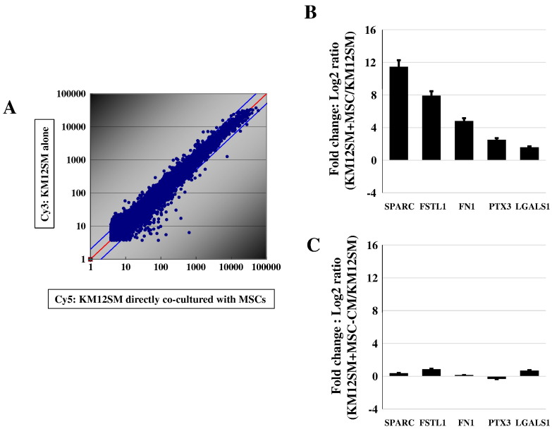
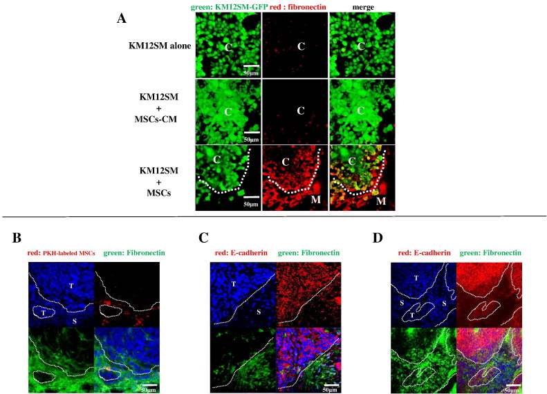
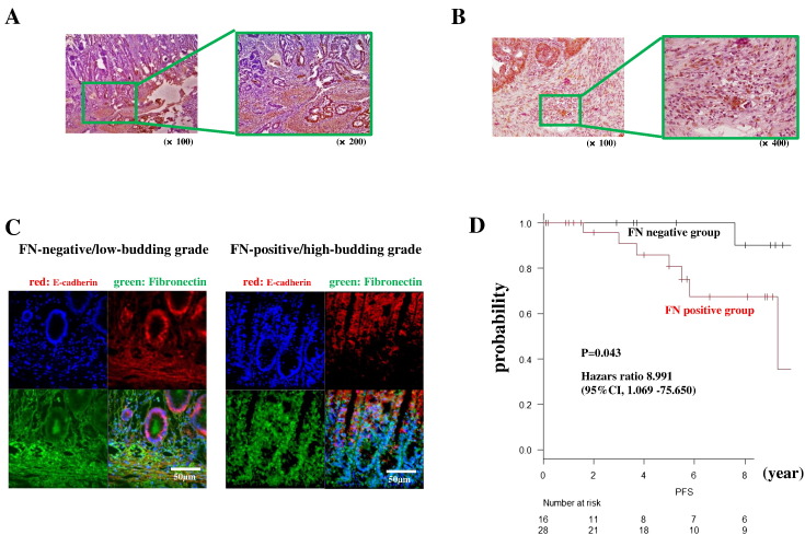
We previously reported that in an orthotopic nude mouse model of human colon cancer, bone marrow-derived mesenchymal stem cells (MSCs) migrated to the tumor stroma and promoted tumor growth and metastasis. Here, we evaluated the proliferation and migration ability of cancer cells cocultured with MSCs to elucidate the mechanism of interaction between cancer cells and MSCs. Proliferation and migration of cancer cells increased following direct coculture with MSCs but not following indirect coculture. Thus, we hypothesized that direct contact between cancer cells and MSCs was important. We performed a microarray analysis of gene expression in KM12SM colon cancer cells directly cocultured with MSCs. Expression of epithelial-mesenchymal transition (EMT)-related genes such as fibronectin (FN), SPARC, and galectin 1 was increased by direct coculture with MSCs. We also confirmed the upregulation of these genes with real-time polymerase chain reaction. Gene expression was not elevated in cancer cells indirectly cocultured with MSCs. Among the EMT-related genes upregulated by direct coculture with MSCs, we examined the immune localization of FN, a well-known EMT marker. In coculture assay in chamber slides, expression of FN was seen only at the edges of cancer clusters where cancer cells directly contacted MSCs. FN expression in cancer cells increased at the tumor periphery and invasive edge in orthotopic nude mouse tumors and human colon cancer tissues. These results suggest that MSCs induce EMT in colon cancer cells via direct cell-to-cell contact and may play an important role in colon cancer metastasis.


Figures

Figure 1
Figure 1
In vitro cell proliferation and migration of KM12SM cells. (A) Proliferation and (B) migration abilities of KM12SM cells cocultured with MSCs compared with those of KM12SM cells cultured alone. (C) Proliferation and (D) migration abilities of KM12SM cells cocultured with MSC-CM (indirect coculture) compared with those of KM12SM cells cultured alone. Time-lapse imaging (obtained with IncuCyte Zoom) of morphology of KM12SM cells cultured (E) alone, (F) with MSCs, or (G) with MSC-CM for 48 hours. Scale bar =100 μm.
Figure 2
Figure 2
Microarray analysis of KM12SM cells. (A) Scatter plots for Cy5-labeled KM12SM cells (directly cocultured with MSCs) and Cy3-labeled KM12SM cells (cultured alone). (B) Expression of SPARC, FSTL1, FN1, PTX3, and LGALS1 in KM12SM cells directly cocultured with MSCs versus expression in KM12SM cells cultured alone, as determined by qRT-PCR. (C) Expression of SPARC, FSTL1, FN1, PTX3, and LGALS1 in KM12SM cells cocultured with MSC-CM versus expression in KM12SM cells cultured alone, as determined by qRT-PCR.
Figure 3
Figure 3
Immunofluorescence staining of KM12SM chamber slides and frozen sections obtained from orthotopic colon cancer models. (A) In vitro expression of FN (red) in KM12SM-GFP colon cancer cells (green) cultured alone (upper panels), with MSC-CM (middle panels), or with MSCs (lower panels). Merged images show regions of overlap. C, KM12SM cancer cells; M, MSCs. Scale bar =50 μm. (B) Expression of FN (green) after injection of PKH26-GL–labeled MSCs (red) into the tail veins of KM12SM tumor-bearing mice. DAPI nuclear staining is shown in blue. Merged image shown in the lower right. T, tumor; S, stroma. Scale bar =50 μm. (C) Expression of FN (green) and E-cadherin (red) in tumors generated by implantation of KM12SM cells only. DAPI nuclear staining is shown in blue. Merged image shown in the lower right. T, tumor; S, stroma. Scale bar =50 μm. (D) Expression of FN (green) and E-cadherin (red) in tumors generated by coimplantation of KM12SM cells and MSCs. DAPI nuclear staining is shown in blue. Merged image shown in the lower right. T, tumor; S, stroma. Scale bar =50 μm.
Figure 4
Figure 4
Expression of FN in surgical specimens of human colon tumors. (A) Expression of FN in the stromal area and at the invasive edges of cancer cells close to the stromal area. (B) Expression of FN in budding cells. (C) Expression of FN (green) and E-cadherin (red) in FN-negative/low–budding grade and FN-positive/high–budding grade patients. DAPI nuclear staining is shown in blue. Merged image shown in lower right. (D) Kaplan-Meier survival curves showing overall survival in FN-positive and FN-negative patient groups.

References

    1. Jemal A, Bray F, Center MM, Ferlay J, Ward E, Forman D. Global cancer statistics. CA Cancer J Clin. 2011;61:69–90. - PubMed
    1. Mantovani A, Allavena P, Sica A, Balkwill F. Cancer-related inflammation. Nature. 2008;454:436–444. - PubMed
    1. Whiteside TL. The tumor microenvironment and its role in promoting tumor growth. Oncogene. 2008;27:5904–5912. - PMC - PubMed
    1. Kitadai Y, Sasaki T, Kuwai T, Nakamura T, Bucana CD, Fidler IJ. Targeting the expression of platelet-derived growth factor receptor by reactive stroma inhibits growth and metastasis of human colon carcinoma. Am J Pathol. 2006;169:2054–2065. - PMC - PubMed
    1. Orimo A, Gupta PB, Sgroi DC, Arenzana-Seisdedos F, Delaunay T, Naeem R, Carey VJ, Richardson AL, Weinberg RA. Stromal fibroblasts present in invasive human breast carcinomas promote tumor growth and angiogenesis through elevated SDF-1/CXCL12 secretion. Cell. 2005;121:335–348. - PubMed