. 2017 Jun:55:100-108.
doi: 10.1016/j.actbio.2017.04.019. Epub 2017 Apr 20.

Regulation of human nucleus pulposus cells by peptide-coupled substrates

Affiliations

Regulation of human nucleus pulposus cells by peptide-coupled substrates

Devin T Bridgen et al. Acta Biomater. 2017 Jun.

Abstract

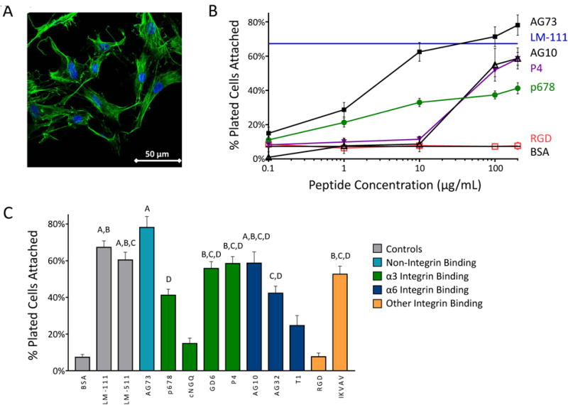
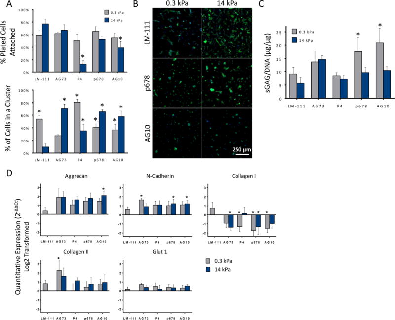
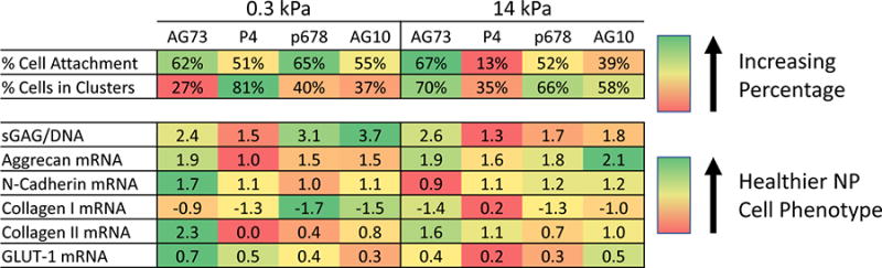
Nucleus pulposus (NP) cells are derived from the notochord and differ from neighboring cells of the intervertebral disc in phenotypic marker expression and morphology. Adult human NP cells lose this phenotype and morphology with age in a pattern that contributes to progressive disc degeneration and pathology. Select laminin-mimetic peptide ligands and substrate stiffnesses were examined for their ability to regulate human NP cell phenotype and biosynthesis through the expression of NP-specific markers aggrecan, N-cadherin, collagen types I and II, and GLUT1. Peptide-conjugated substrates demonstrated an ability to promote expression of healthy NP-specific markers, as well as increased biosynthetic activity. We show an ability to re-express markers of the juvenile NP cell and morphology through control of peptide presentation and stiffness on well-characterized polyacrylamide substrates. NP cells cultured on surfaces conjugated with α3 integrin receptor peptides P4 and P678, and on α2, α5, α6, β1 integrin-recognizing peptide AG10, show increased expression of aggrecan, N-cadherin, and types I and II collagen, suggesting a healthier, more juvenile-like phenotype. Multi-cell cluster formation was also observed to be more prominent on peptide-conjugated substrates. These findings indicate a critical role for cell-matrix interactions with specific ECM-mimetic peptides in supporting and maintaining a healthy NP cell phenotype and bioactivity.

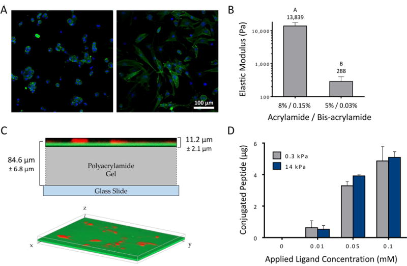
Statement of significance: NP cells reside in a laminin-rich environment that deteriorates with age, including a loss of water content and changes in the extracellular matrix (ECM) structure that may lead to the development of a degenerated IVD. There is great interest in methods to re-express healthy, biosynthetically active NP cells using laminin-derived biomimetic peptides toward the goal of using autologous cell sources for tissue regeneration. Here, we describe a novel study utilizing several laminin mimetic peptides conjugated to polyacrylamide gels that are able to support an immature, healthy NP phenotype after culture on "soft" peptide gels. These findings can support future studies in tissue regeneration where cells may be directed to a desired regenerative phenotype using niche-specific ECM peptides.

Keywords: Cell-matrix interactions; Extracellular matrix; Hydrogel; Mechanobiology; Polyacrylamide; Spine.


Figures

Figure 1
Figure 1
(A) Primary human NP cells display consistent morphology and higher attachment on LM-111-coated tissue culture plastic (scale bar = 50 μm). (B) NP cells attach to peptide-coated surfaces at levels equivalent to that for LM-111, but only at higher peptide densities and for select peptides as shown here. (C) Cell numbers attaching to peptide coated surfaces for all screened peptides at 200 μg/mL. Conditions labeled with the same letter (A, B, etc) are equivalent and significantly different than the BSA control (p<0.05, ANOVA with post-hoc tests). SEM are shown.
Figure 2
Figure 2
(A) Primary human NP cells attach to soft LM-111 coupled substrates (0.3 kPa polyacrylamide; left image) as rounded cells forming multi-cell clusters, while cells attaching to stiff (14 kPa; right image) substrates may elongate and form fewer cell clusters. (B) PAAm gels were formed with varying bisacrylamide ratios to achieve compressive moduli of 0.3 kPa (soft) and 14 kPa (stiff) as measured by atomic force microscopy. Conditions not connected by the same letters are significantly different from each other (p<0.0001, Mann-Whitney test). SD are shown. (C) Representative confocal Z-stack images of fluorescently labeled peptides conjugated to polyacrylamide gel. Peptides (green, FITC) and cells (red, mCherry) are shown in relation to each other, superimposed over a scaled diagram of the polyacrylamide gel (top). A three-dimensional isometric confocal image illustrates the even distribution of peptides throughout the gel (bottom). (D) Peptide functionalized substrates were found to have equivalent peptide densities on soft and stiff gels, with differences that vary with the amount of applied peptide. All studies were performed at the highest degree of peptide functionalization, 0.1 mM. SEM are shown.
Figure 3
Figure 3
(A) Primary human NP cells attached to both soft and stiff peptide functionalized acrylamide gels. All peptide functionalized gels promoted greater cell clustering than for stiff LM-111 functionalized substrates (* p<0.05, significantly different from stiff LM-111 functionalized gels) SEM are shown. (B) Representative day 4 images showing the effect of substrate stiffness and ligand for NP cell morphologies. A majority of attached cells interact to form rounded, multi-cell clusters, in comparison to cells on the stiff (14 kPa) LM-111 control showing flattened morphology with stress fiber formation (scale bar = 250 μm). (C) NP cell production of sGAG per DNA varied across peptide-functionalized substrates. As found previously, sGAG/DNA for NP cells upon soft LM-111-functionalized substrates was greater (1.6-fold) than for stiff substrates. While similar elevations in sGAG/DNA were observed for many peptide-functionalized gels, only soft gels functionalized with P678 and AG10 were associated with higher sGAG/DNA (* p<0.05, significantly greater than stiff LM-111, 2-way ANOVA, Dunnett's post hoc). SEM are shown. (D) mRNA levels for aggrecan (AGC), N-cadherin (CDH2), type I collagen (COLA1), type II collagen (COL2A1), and GLUT-1 in primary human NP cells. (*p<0.05, significantly different from stiff LM-111 functionalized polyacrylamide gels, Dunnett's post hoc). SEM are shown.
Figure 4
Figure 4
Heat map illustrating average values within each variable for each peptide functionalized substrates. Green denotes values closer to those recognized as healthy NP-specific. Each row demonstrates the best condition for each parameter, but is not compared to different items (i.e. other rows). As an example, NP cells with higher sGAG synthesis and lower COL1A1 mRNA may be produced by periods of culture upon soft substrates functionalized with P678 or AG10.

References

    1. Sakai D, Nakamura Y, Nakai T, Mishima T, Kato S, Grad S, Alini M, Risbud MV, Chan D, Cheah KS, Yamamura K, Masuda K, Okano H, Ando K, Mochida J. Exhaustion of nucleus pulposus progenitor cells with ageing and degeneration of the intervertebral disc. Nature communications. 2012;3:1264. - PMC - PubMed
    1. Risbud MV, Schoepflin ZR, Mwale F, Kandel RA, Grad S, Iatridis JC, Sakai D, Hoyland JA. Defining the phenotype of young healthy nucleus pulposus cells: recommendations of the Spine Research Interest Group at the 2014 annual ORS meeting. J Orthop Res. 2015;33(3):283–93. - PMC - PubMed
    1. Choi KS, Harfe BD. Hedgehog signaling is required for formation of the notochord sheath and patterning of nuclei pulposi within the intervertebral discs. Proceedings of the National Academy of Sciences of the United States of America. 2011;108(23):9484–9. - PMC - PubMed
    1. Tran CM, Schoepflin ZR, Markova DZ, Kepler CK, Anderson DG, Shapiro IM, Risbud MV. CCN2 suppresses catabolic effects of interleukin-1beta through alpha5beta1 and alphaVbeta3 integrins in nucleus pulposus cells: implications in intervertebral disc degeneration. The Journal of biological chemistry. 2014;289(11):7374–87. - PMC - PubMed
    1. Risbud MV, Guttapalli A, Albert TJ, Shapiro IM. Hypoxia activates MAPK activity in rat nucleus pulposus cells: regulation of integrin expression and cell survival. Spine (Phila Pa 1976) 2005;30(22):2503–9. - PubMed

Publication types

MeSH terms