

Establishment and characterization of novel epithelial-like cell lines derived from human periodontal ligament tissue in vitro
- PMID: 28434170
- PMCID: PMC5646140
- DOI: 10.1007/s13577-017-0173-y
Establishment and characterization of novel epithelial-like cell lines derived from human periodontal ligament tissue in vitro
Abstract
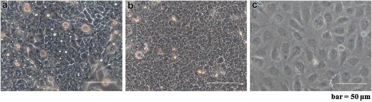
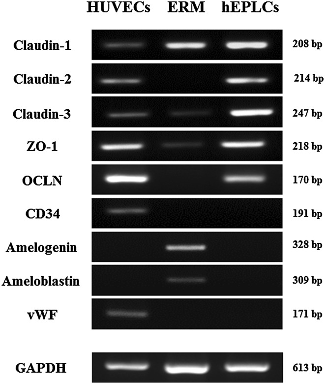
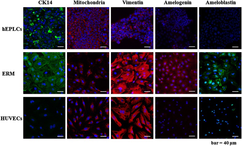
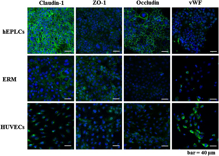
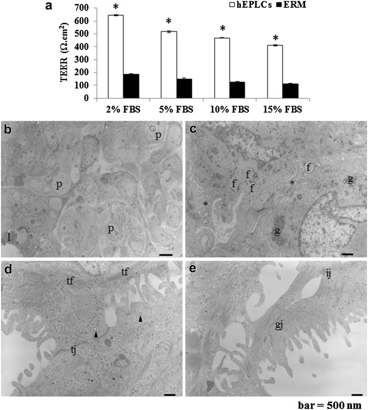
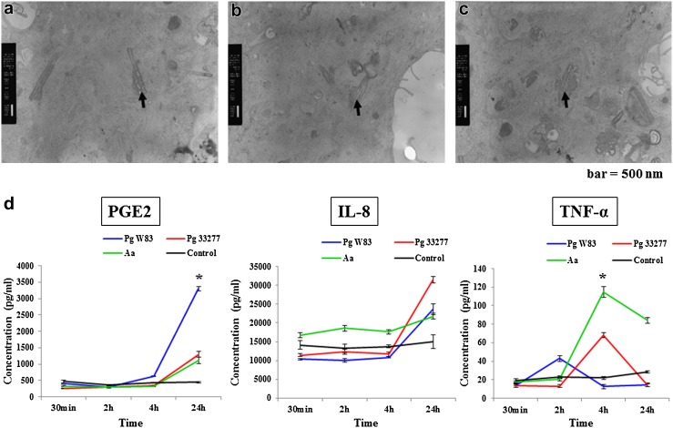
In this study, novel human-derived epithelial-like cells (hEPLCs) lines were established from periodontal ligament (PDL) tissues, which were composed of a variety of cell types and exhibited complex cellular activities. To elucidate the putative features distinguishing these from epithelial rest of Malassez (ERM), we characterized hEPLCs based on cell lineage markers and tight junction protein expression. The aim of this study was, therefore, to establish and characterize hEPLCs lines from PDL tissues. The hEPLCs were isolated from PDL of third molar teeth. Cellular morphology and cell organelles were observed thoroughly. The characteristics of epithelial-endothelial-mesenchymal-like cells were compared in several markers by gene expression and immunofluorescence, to ERM and human umbilical-vein endothelial cells (HUVECs). The resistance between cellular junctions was assessed by transepithelial electron resistance, and inflammatory cytokines were detected by ELISA after infecting hEPLCs with periodontopathic bacteria. The hEPLCs developed into small epithelial-like cells in pavement appearance similar to ERM. However, gene expression patterns and immunofluorescence results were different from ERM and HUVECs, especially in tight junction markers (Claudin, ZO-1, and Occludins), and endothelial markers (vWF, CD34). The transepithelial electron resistance indicated higher resistance in hEPLCs, as compared to ERM. Periodontopathic bacteria were phagocytosed with upregulation of inflammatory cytokine secretion within 24 h. In conclusion, hEPLCs that were derived using the single cell isolation method formed tight multilayers colonies, as well as strongly expressed tight junction markers in gene expression and immunofluorescence. Novel hEPLCs lines exhibited differently from ERM, which might provide some specific functions such as metabolic exchange and defense mechanism against bacterial invasion in periodontal tissue.
Keywords: EMT; Epithelial rests of Malassez differentiation; Epithelial-like cells; Establishment cell lines; Periodontal ligament tissue.
Conflict of interest statement
Conflict of interest
The authors declare that they have no competing interests.
Ethical approval
The protocol performed in this study involving human participants were approved by the ethics committee of Nippon Dental University.
Informed consent
Informed consent was obtained from all individual participants included in the study.
Figures
Similar articles
-
In vitro characterization of the cytokine profile of the epithelial cell rests of Malassez.J Periodontol. 2008 May;79(5):912-9. doi: 10.1902/jop.2008.070553. J Periodontol. 2008. PMID: 18454671
-
Induction of Periodontal Ligament-like Cells by Coculture of Dental Pulp Cells, Dedifferentiated Cells Generated from Epithelial Cell Rests of Malassez, and Umbilical Vein Endothelial Cells.J Endod. 2022 Nov;48(11):1387-1394. doi: 10.1016/j.joen.2022.08.006. Epub 2022 Sep 5. J Endod. 2022. PMID: 36067833
-
Epithelial cell rests of Malassez contain unique stem cell populations capable of undergoing epithelial-mesenchymal transition.Stem Cells Dev. 2012 Jul 20;21(11):2012-25. doi: 10.1089/scd.2011.0471. Epub 2012 Jan 26. Stem Cells Dev. 2012. PMID: 22122577 Free PMC article.
-
Role of the epithelial cell rests of Malassez in the development, maintenance and regeneration of periodontal ligament tissues.Periodontol 2000. 2013 Oct;63(1):217-33. doi: 10.1111/prd.12023. Periodontol 2000. 2013. PMID: 23931062 Review.
-
The epithelial cell rests of Malassez--a role in periodontal regeneration?J Periodontal Res. 2006 Aug;41(4):245-52. doi: 10.1111/j.1600-0765.2006.00880.x. J Periodontal Res. 2006. PMID: 16827716 Review.
Cited by
-
Mechanical stress alters protein O-GlcNAc in human periodontal ligament cells.J Cell Mol Med. 2019 Sep;23(9):6251-6259. doi: 10.1111/jcmm.14509. Epub 2019 Jun 25. J Cell Mol Med. 2019. PMID: 31237748 Free PMC article.
References
-
- Beertsen W, McCulloch CAG, Sodek J. The periodontal ligament: a unique, multifunctional connective tissue. Periodontology. 2000;1997(13):20–40. - PubMed
-
- Nanci A. Development of the tooth and its supporting tissues. Ten Cate’s oral histology: development, structure, and function. 8. St. Louis: Elsevier; 2013. pp. 70–94.
MeSH terms
Substances
Grants and funding
LinkOut - more resources
Full Text Sources
Other Literature Sources
Miscellaneous
