. 2017 May 4;169(4):636-650.e14.
doi: 10.1016/j.cell.2017.03.042. Epub 2017 Apr 20.

Stem Cell Lineage Infidelity Drives Wound Repair and Cancer

Affiliations

Stem Cell Lineage Infidelity Drives Wound Repair and Cancer

Yejing Ge et al. Cell. .

Abstract

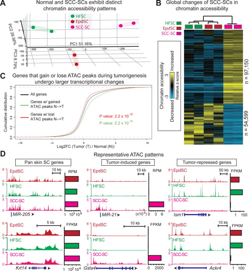
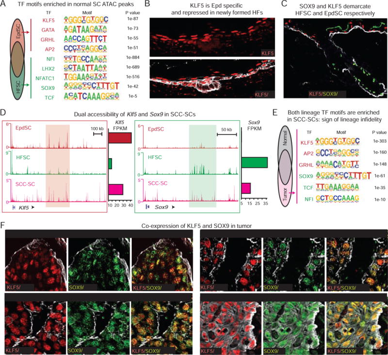
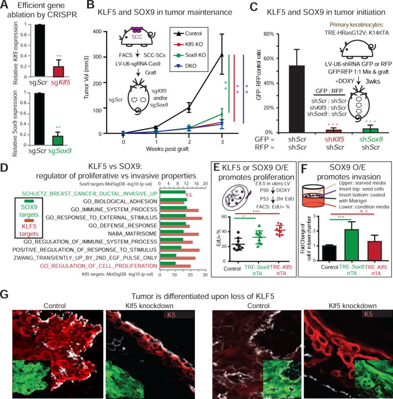
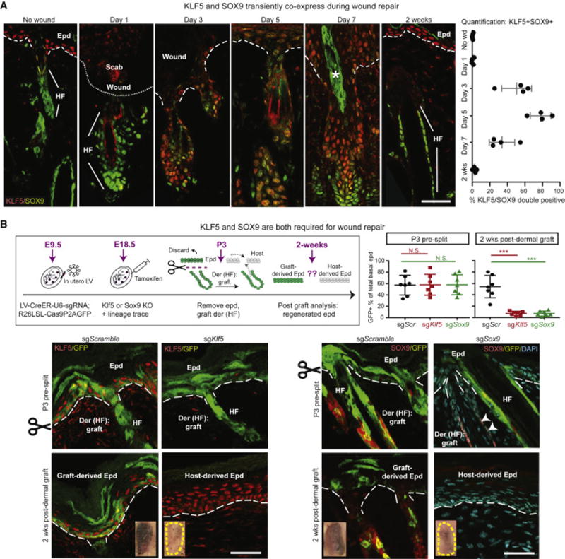
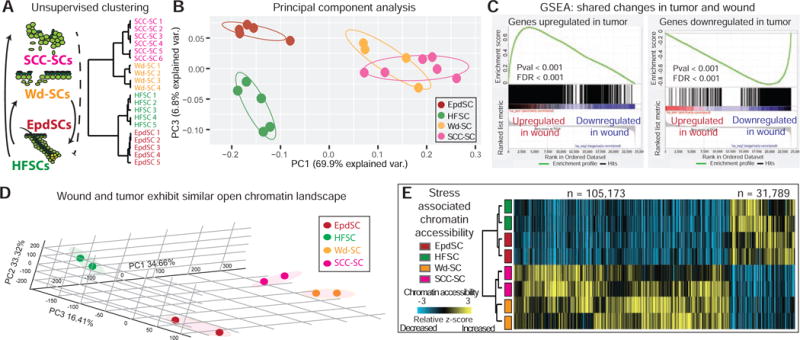
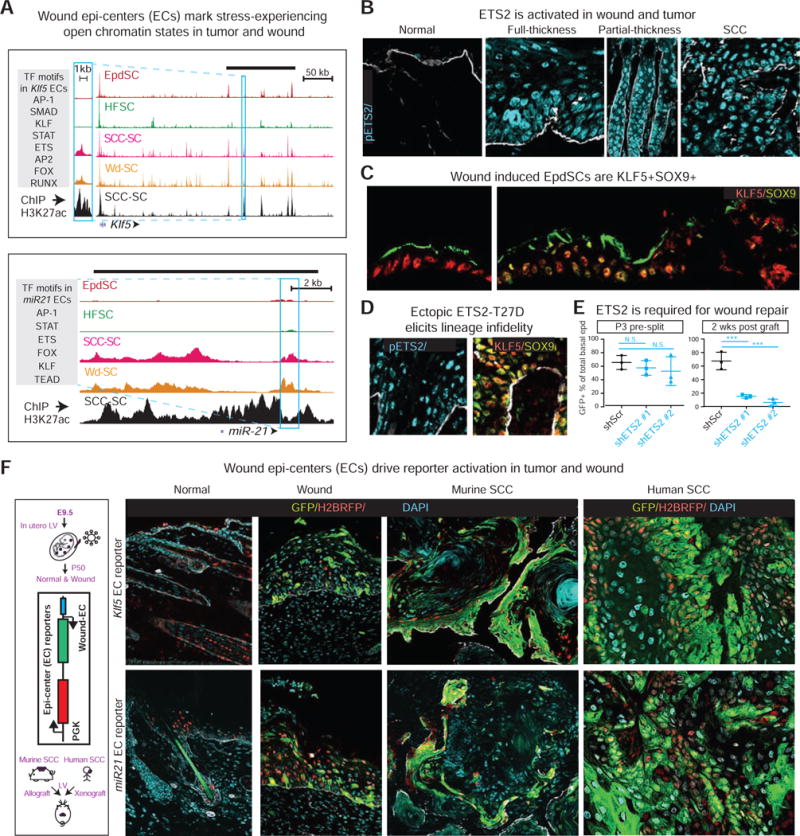
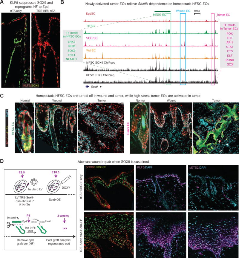
Tissue stem cells contribute to tissue regeneration and wound repair through cellular programs that can be hijacked by cancer cells. Here, we investigate such a phenomenon in skin, where during homeostasis, stem cells of the epidermis and hair follicle fuel their respective tissues. We find that breakdown of stem cell lineage confinement-granting privileges associated with both fates-is not only hallmark but also functional in cancer development. We show that lineage plasticity is critical in wound repair, where it operates transiently to redirect fates. Investigating mechanism, we discover that irrespective of cellular origin, lineage infidelity occurs in wounding when stress-responsive enhancers become activated and override homeostatic enhancers that govern lineage specificity. In cancer, stress-responsive transcription factor levels rise, causing lineage commanders to reach excess. When lineage and stress factors collaborate, they activate oncogenic enhancers that distinguish cancers from wounds.

Keywords: cancer; epigenetics; lineage infidelity; regeneration; skin; stem cells; stress response; super-enhancers; transcriptional regulation; wound repair.


Figures

Figure 1
Figure 1. Tumor Stem Cells Undergo Global Changes in Chromatin Accessibility Compared to Their Normal Counterparts
(A) Principal component analysis of ATACseq signals from biological duplicates of FACS sorted tumor (SCC-) and normal (HF- and Epd-) SCs. (B) Genome-wide ATAC signals in SCC-, Epd- and HF-SCs are z-score-transformed and averaged across 100-bp genomic windows (n=# windows plotted). Hierarchical clustering shows comparison of open chromatin regions between normal (Epd and HF) and SCC-SCs. Heatmap shows gain (yellow), loss (blue) or no change (black) of ATAC signals from normal to stress comparison. (C) Cumulative density plot shows that genes upregulated in tumor (T) versus normal (N) SCs have gained ATAC peaks (log2FC positive, green curve right shift), whereas those downregulated have lost ATAC peaks (log2FC negative, red curve left shift). KS one-sided test was used to compare genes that had gained or lost ATAC peaks in T vs N SCs relative to all genes. (D) SCC-, Epd- and HF-SC ATAC tracks of representative genes that are constitutively active, induced or suppressed in tumors, respectively. Shown to the right are transcript levels (RPM for miRNA reads, FPKM for mRNA reads). Also see Figure S1.
Figure 2
Figure 2. SCC Stem Cells Express Both Epidermal and Hair Follicle Lineage Markers
All images are representative and from at least 5 biologically independent replicates. Bars = 50μm. Dashed line denotes epidermal-dermal border. (A) Motif analysis identifies enriched TF motifs associated with ATAC-peaks unique to HFSCs or EpdSCs compared to peaks common to both lineages. (B) Immunofluorescence reveals downregulation of Epd KLF5 (arrows) in newly emerged hair germs (open arrowheads) at embryonic day E17.5. (C) Lineage restricted SOX9 and KLF5 expression during skin homeostasis. Asterisk denotes artifactual autofluorescence of Epd squames. (D) ATAC tracks reveal enhanced chromatin accessibility within Klf5 and Sox9 regulatory regions (red and green shades) in SCC-SCs compared to SCs of the opposite lineage. mRNA levels (FPKM) at right. (E) Motif analysis identifies enriched TF motifs associated with SCC-SC-exclusive ATAC-peaks compared to those shared between SCC- and normal SCs. (F) Co-expression of SOX9 and KLF5 (arrows) in papillomas, murine and human SCCs, and lung metastases. Also see Figure S2.
Figure 3
Figure 3. Co-expression of KLF5 and SOX9 Is a Functional Hallmark of Tumorigenesis
(A–B) SCCs-SCs were FACS-purified and infected with lentivirus (LV) harboring CAS9 and small guide RNAs against Klf5 (red) or Sox9 (green) or Scrambled (Scr) control (black). Following 2d puromycin selection, cells were subjected to either qPCR (A), or engrafted onto backskins of Nude mice (B). Tumor volume was measured each week thereafter (n=5). (C) Primary keratinocytes from TRE-HRasG12V; K14rtTA mice were transduced with LV-H2BGFP harboring small hairpins against Klf5, Sox9 or Scr. Cells were sorted 3d later, mixed 1:1 GFP/RFP, and engrafted onto backskins of Nude mice with doxycycline. GFP/RFP ratio was measured 3 wks later by FACS. (D) Genes associated with SCC-SC ATAC peaks harboring SOX9 or KLF5 motifs were analyzed by MolSigDB for their associated molecular pathways. Note preference of KLF5 for proliferation and SOX9 for invasion pathway genes. (E) Overexpression (O/E) of KLF5 in vivo was achieved by LV-TRE-Klf5 infection of E9.5 K14-rtTA embryos. O/E of SOX9 in vivo was achieved by LV-rtTA infection of E9.5 TRE-Sox9 embryos. At P50, mice were treated 3d with doxycycline 3d and then pulsed with EdU 2 hr prior to FACS. Shown are % EdU+ of total basal cells. n = 9. (F) Keratinocytes from K14-rtTA mice were infected with LV TRE-Sox9 or TRE-Klf5 (TRE-empty as control) and treated 3d with doxycycline prior to Boyden chamber assays. Keratinocytes in starvation media in the upper chamber were assayed for their ability to invade through matrigel-coated filters to reach the stimulatory dermal fibroblast-conditioned media in the lower chamber. 1d after seeding, cells reaching the bottom were counted, and results were plotted as fold change compared to control. n = 6. (G) Immunofluorescence of engrafted SCC-SCs (LV-GFP transduced to mark tumor cells) knocked down for Klf5. Note decrease in progenitor marker K5 and increase in differentiation marker K10. Bar = 50μm. At least 5 biologically independent replicates were analyzed. Shown are representative images. Paired t tests were performed for A, C, E, F. Two-way ANOVA with repeated measurement was performed for B. Shown as mean ± std. *P <0.05, **P<0.01. ***P <0.001. N.S. not significant. Also see Figure S3, Table S1 and S2.
Figure 4
Figure 4. Epithelial Wounding Transiently Inflicts and Relies on Lineage Infidelity for Repair
All images are representative and from at least 3 biologically independent replicates. Bars = 50μm. Dashed line denotes epidermal-dermal border. (A) Temporal changes of SOX9 and KLF5 following a partial-thickness wound. Note co-expression peaks at height of wound-repair, and is subsequently resolved. Asterisk denotes artifactual autofluorescence of hair shaft. (B) (Top left) Experimental design for inducing epithelial-specific Klf5 and Sox9 knockout by in vivo CRISPR/CAS and analyzing consequences to wound-repair. R26-LSL-Cas9-P2A-GFP E9.5 embryos were transduced with LV-CreER-U6-sgKlf5 or -sgSox9, followed by tamoxifen at E18.5. Postnatal pup backskin was either directly frozen for sectioning and immunofluorescence, or treated with EDTA to remove epidermis prior to dermal grafting. Grafts were analyzed 2wk later by FACS quantification of % GFP+ cells within total re-epithelialized EpdSCs (top right), or by immunofluorescence (bottom row). Note that in contrast to Scr sg controls, no contribution from GFP+ HF cells is detected in the regenerated epidermis without either Klf5 or Sox9. Arrowheads point to SOX9 signal in GFP- cells. n = 7 for FACS quantification. Paired t test was performed. ***P<0.001. N.S. not significant. Also see Figure S4.
Figure 5
Figure 5. Wounded and Tumorigenic Stem Cells Display Similar Transcriptomes and Genome-wide Chromatin Accessibilities
(A–B) Unsupervised hierarchical clustering and principal component analysis (PCA) of RNAseq data reveals similarities between SCC- and wounded (Wd)-SCs relative to homeostatic SCs. (C) Gene set enrichment analysis (GSEA) reveals striking parallels in transcriptome changes that occur in tumor and wound vs normal SCs. Gene changes in tumor vs normal are compared against pre-ranked changes in wound vs normal. (D) PCA of ATACseq signals from biological duplicates of FACS sorted tumor (SCC-), wound (Wd-), and normal (HF and Epd) SCs. (E) Genome-wide ATAC signals in Wd-, SCC-, Epd- and HF-SCs are z-score-transformed and averaged across 100-bp genomic windows (N=# windows plotted). Hierarchical clustering shows divergence between normal (Epd and HF) and stress-experienced (Wd and SCC) SCs. Heatmap shows gain (yellow), loss (blue) or no change (black) of ATAC signals in normal:stress comparisons. See also Figure S5.
Figure 6
Figure 6. A Role for Stress-Induced Transcription Factors in Driving Lineage Infidelity in Wounds and Tumors
All fluorescence images of tissues are representative of at least 3 biologically independent replicates. Bars = 50μm. Dashed line denotes epidermal-dermal border. Asterisks denote artifactual autofluorescence of Epd squames (C) or hair shaft (F). (A) Shown are Klf5 and miR-221 ATAC of Wd-, SCC-, Epd- and HF-SCs and H3K27Ac ChIPseq of SCC-SCs. Boxes denote epicenters (ECs) cloned for testing their enhancer activities with eGFP reporters in vivo. Grey shaded boxes list TF motifs within these ECs. Overhead black bars denote annotated super-enhancers (SE) for Klf5 and miR-221 in SCC-SCs (Yang et al., 2015). (B) pETS2 is induced in tumor and wound. (C) SOX9 is induced in KLF5+ wounded epidermis (Epd), indicating that lineage infidelity occurs in Epd as well upon injury. Arrows indicate direction of wound closure. (D) Forced activation of ETS2 in skin epithelium by transducing K14rtTA embryos with LV TRE-T72D-Ets2, and followed by 4wks doxycycline starting at P0 (Yang et al., 2015). Backskin was analyzed for pETS2, KLF5 and SOX9. Note ectopic KLF5 in HF and SOX9 in Epd. (E) Ets2 knockdown in vivo dramatically impairs the ability of SCs to contribute to wound-repair. Shown are quantifications (n=3) of re-epithelialization contribution when split thickness grafts are deficient for ETS2 with two different shRNAs. (F) Wound-induced epicenter-driven reporter activity (eGFP), in tumors and wounds, but not homeostatic skin. Control PGK-H2BRFP reveals equivalent LV transduction in all states. See also Figure S6.
Figure 7
Figure 7. Newly activated epicenters Lock Tumors Into Sustained Lineage Infidelity
All fluorescence images are representative and from at least 3 biologically independent replicates. Bars = 50μm. Dashed line denotes epidermal-dermal border. (A) Within 2 wks forced activation of KLF5, SOX9 is suppressed in adult HFs. (B) ATAC peaks at the Sox9 locus of Wd-, SCC-, Epd- and HF-SCs, and contrasted with SOX9 and LHX2 ChIPseq peaks of HFSC-SCs. Three types of epicenters are boxed: HFSC-EC green, Tumor-EC pink, Wound-EC blue. Note silencing of Sox9’s homeostatic enhancer and activation of new tumor-specific enhancer in SCC-SCs. (C) Sox9’s homeostatic enhancer drives GFP reporter activity only in HFSCs and not in tumor or wound states, while Sox9’s tumor-EC is active only in SCCs and not in wounds or normal homeostatic skin. Note the contrast to wound-ECs, which are active in both wounds and tumors (Figure 6). All reporters were transduced comparably (H2BRFP). (D) Forced activation of SOX9 in skin epithelium was achieved by transducing TRE-Sox9 embryos with LV rtTA-H2BGFP, and then treating animals with doxycycline just prior to split-thickness engraftment. Skin grafts were then analyzed 2wk later, with half used for sectioning and immunofluorescence and the other half for EdU pulse and analyses. See also Figure S7.

Comment in

Similar articles

Cited by

References

    1. Adam RC, Yang H, Rockowitz S, Larsen SB, Nikolova M, Oristian DS, Polak L, Kadaja M, Asare A, Zheng D, et al. Pioneer factors govern super-enhancer dynamics in stem cell plasticity and lineage choice. Nature. 2015;521:366–370. - PMC - PubMed
    1. Akiyama H, Kim JE, Nakashima K, Balmes G, Iwai N, Deng JM, Zhang Z, Martin JF, Behringer RR, Nakamura T, et al. Osteo-chondroprogenitor cells are derived from Sox9 expressing precursors. Proc Natl Acad Sci U S A. 2005;102:14665–14670. - PMC - PubMed
    1. Antsiferova M, Werner S. The bright and the dark sides of activin in wound healing and cancer. J Cell Sci. 2012;125:3929–3937. - PubMed
    1. Arwert EN, Hoste E, Watt FM. Epithelial stem cells, wound healing and cancer. Nat Rev Cancer. 2012;12:170–180. - PubMed
    1. Balmain A, Yuspa SH. Milestones in skin carcinogenesis: the biology of multistage carcinogenesis. J Invest Dermatol. 2014;134:E2–7. - PubMed