

Transfer of C-terminal residues of human apolipoprotein A-I to insect apolipophorin III creates a two-domain chimeric protein with enhanced lipid binding activity
- PMID: 28434970
- PMCID: PMC5518692
- DOI: 10.1016/j.bbamem.2017.04.017
Transfer of C-terminal residues of human apolipoprotein A-I to insect apolipophorin III creates a two-domain chimeric protein with enhanced lipid binding activity
Abstract
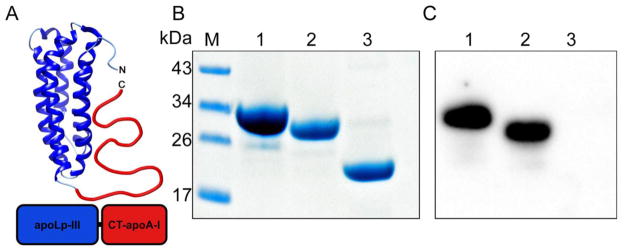
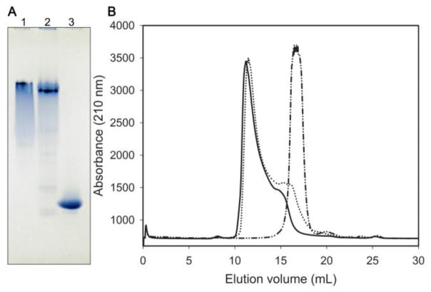
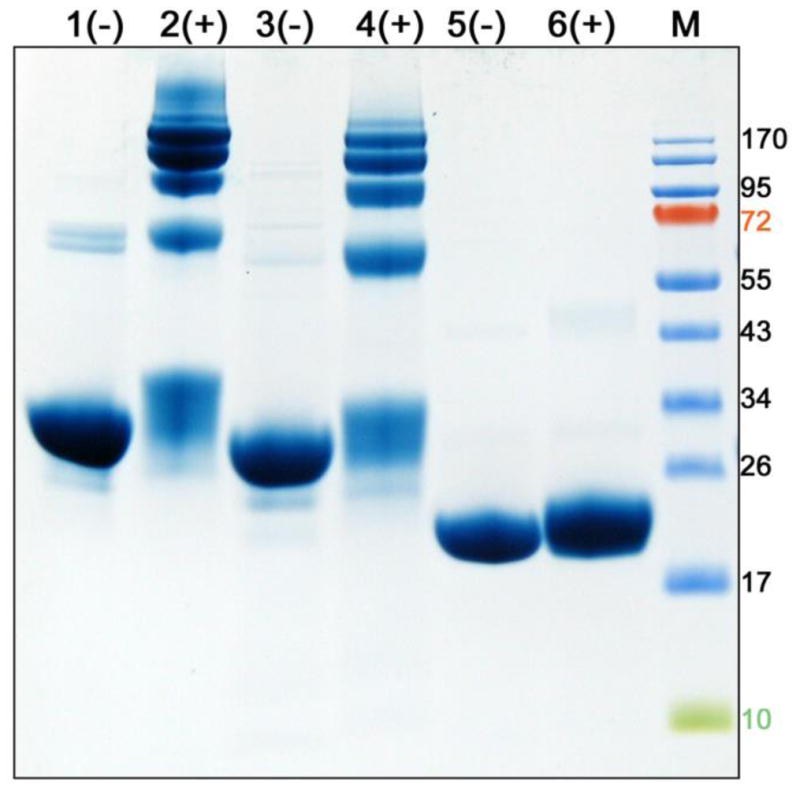
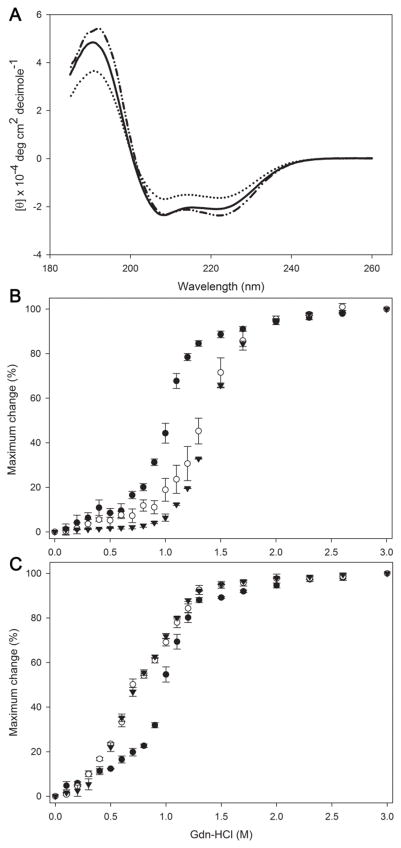
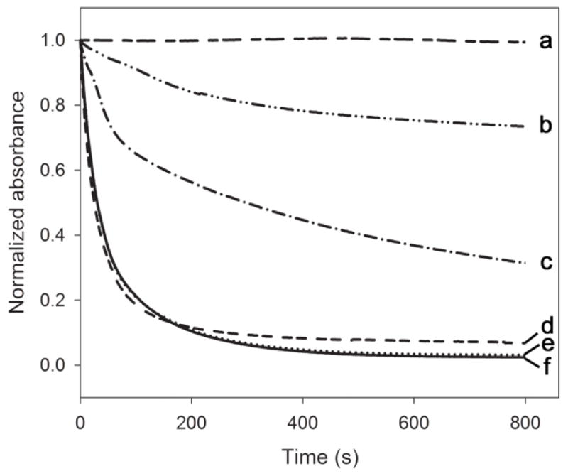
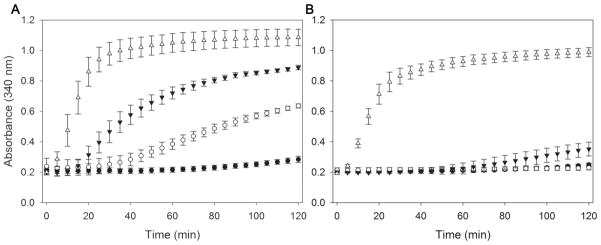
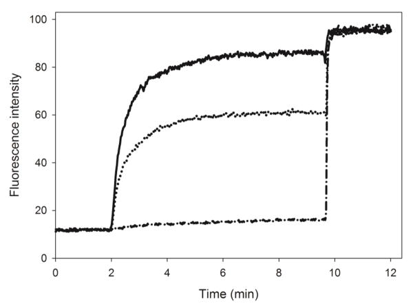
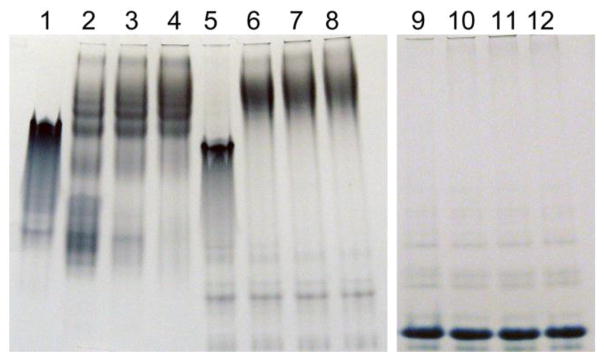
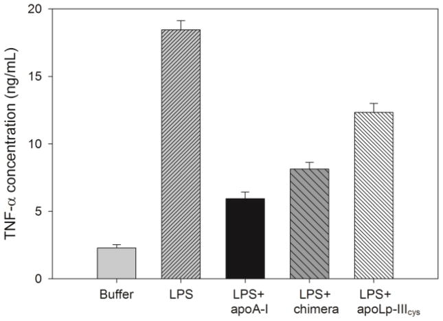
Apolipophorin III (apoLp-III) is an insect apolipoprotein (18kDa) that comprises a single five-helix bundle domain. In contrast, human apolipoprotein A-I (apoA-I) is a 28kDa two-domain protein: an α-helical N-terminal domain (residues 1-189) and a less structured C-terminal domain (residues 190-243). To better understand the apolipoprotein domain organization, a novel chimeric protein was engineered by attaching residues 179 to 243 of apoA-I to the C-terminal end of apoLp-III. The apoLp-III/apoA-I chimera was successfully expressed and purified in E. coli. Western blot analysis and mass spectrometry confirmed the presence of the C-terminal domain of apoA-I within the chimera. While parent apoLp-III did not self-associate, the chimera formed oligomers similar to apoA-I. The chimera displayed a lower α-helical content, but the stability remained similar compared to apoLp-III, consistent with the addition of a less structured domain. The chimera was able to solubilize phospholipid vesicles at a significantly higher rate compared to apoLp-III, approaching that of apoA-I. The chimera was more effective in protecting phospholipase C-treated low density lipoprotein from aggregation compared to apoLp-III. In addition, binding interaction of the chimera with phosphatidylglycerol vesicles and lipopolysaccharides was considerably improved compared to apoLp-III. Thus, addition of the C-terminal domain of apoA-I to apoLp-III created a two-domain protein, with self-association, lipid and lipopolysaccharide binding properties similar to apoA-I. The apoA-I like behavior of the chimera indicate that these properties are independent from residues residing in the N-terminal domain of apoA-I, and that they can be transferred from apoA-I to apoLp-III.
Keywords: Apolipophorin III; Apolipoprotein; Lipid binding; Lipoprotein.
Copyright © 2017 Elsevier B.V. All rights reserved.
Figures
Similar articles
-
Insights into the C-terminal domain of apolipoprotein E from chimera studies with apolipophorin III.Mol Cell Biochem. 2023 Jan;478(1):173-183. doi: 10.1007/s11010-022-04497-y. Epub 2022 Jun 28. Mol Cell Biochem. 2023. PMID: 35763125 Free PMC article.
-
Expression of the C-terminal domain of human apolipoprotein A-I using a chimeric apolipoprotein.Protein Expr Purif. 2017 Sep;137:13-19. doi: 10.1016/j.pep.2017.06.008. Epub 2017 Jun 15. Protein Expr Purif. 2017. PMID: 28624493 Free PMC article.
-
An N-terminal three-helix fragment of the exchangeable insect apolipoprotein apolipophorin III conserves the lipid binding properties of wild-type protein.Biochemistry. 2001 Mar 13;40(10):3150-7. doi: 10.1021/bi0013804. Biochemistry. 2001. PMID: 11258930
-
Merck Frosst award lecture 1995. La conference Merck Frosst 1995. Structural studies of lipoproteins and their apolipoprotein components.Biochem Cell Biol. 1996;74(2):155-64. doi: 10.1139/o96-016. Biochem Cell Biol. 1996. PMID: 9213424 Review.
-
Apolipophorin III: role model apolipoprotein.Insect Biochem Mol Biol. 2006 Apr;36(4):231-40. doi: 10.1016/j.ibmb.2006.01.001. Epub 2006 Jan 18. Insect Biochem Mol Biol. 2006. PMID: 16551537 Review.
Cited by
-
The role of C-terminal ionic residues in self-association of apolipoprotein A-I.Biochim Biophys Acta Biomembr. 2023 Feb;1865(2):184098. doi: 10.1016/j.bbamem.2022.184098. Epub 2022 Dec 6. Biochim Biophys Acta Biomembr. 2023. PMID: 36481181 Free PMC article.
-
Isoform-specific modification of apolipoprotein E by 4-hydroxynonenal: protective role of apolipoprotein E3 against oxidative species.FEBS J. 2023 Jun;290(11):3006-3025. doi: 10.1111/febs.16729. Epub 2023 Feb 8. FEBS J. 2023. PMID: 36661393 Free PMC article.
-
Charged Residues in the C-Terminal Domain of Apolipoprotein A-I Modulate Oligomerization.Biochemistry. 2018 Apr 17;57(15):2200-2210. doi: 10.1021/acs.biochem.7b01052. Epub 2018 Apr 3. Biochemistry. 2018. PMID: 29578333 Free PMC article.
-
Insights into the C-terminal domain of apolipoprotein E from chimera studies with apolipophorin III.Mol Cell Biochem. 2023 Jan;478(1):173-183. doi: 10.1007/s11010-022-04497-y. Epub 2022 Jun 28. Mol Cell Biochem. 2023. PMID: 35763125 Free PMC article.
-
Expression of the C-terminal domain of human apolipoprotein A-I using a chimeric apolipoprotein.Protein Expr Purif. 2017 Sep;137:13-19. doi: 10.1016/j.pep.2017.06.008. Epub 2017 Jun 15. Protein Expr Purif. 2017. PMID: 28624493 Free PMC article.
References
-
- Jonas A, Phillips MC. Lipoprotein structure. In: Vance DE, Vance JE, editors. Biochemistry of Lipids, Lipoproteins and Membranes. Elsevier BV; Amsterdam: 2008. pp. 485–506.
-
- Saito H, Lund-Katz S, Phillips MC. Contributions of domain structure and lipid interaction to the functionality of exchangeable human apolipoproteins. Prog Lipid Res. 2004;43:350–380. - PubMed
Publication types
MeSH terms
Substances
Grants and funding
LinkOut - more resources
Full Text Sources
Other Literature Sources
Miscellaneous
