

Methotrexate-coupled nanoparticles and magnetic nanochemothermia for the relapse-free treatment of T24 bladder tumors
- PMID: 28435259
- PMCID: PMC5388224
- DOI: 10.2147/IJN.S120969
Methotrexate-coupled nanoparticles and magnetic nanochemothermia for the relapse-free treatment of T24 bladder tumors
Abstract
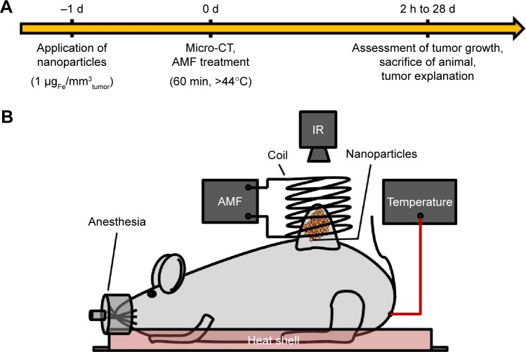
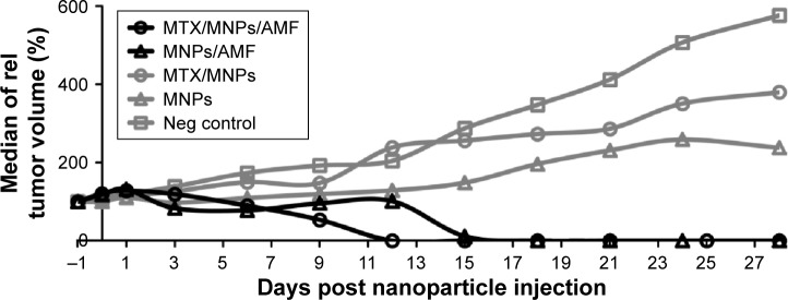
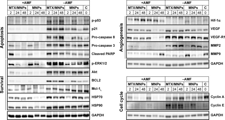
Heat-based approaches have been considered as promising tools due to their ability to directly eradicate tumor cells and/or increase the sensitivity of tumors to radiation- or chemotherapy. In particular, the heating of magnetic nanoparticles (MNPs) via an alternating magnetic field can provide a handy alternative for a localized tumor treatment. To amplify the efficacy of magnetically induced thermal treatments, we elucidated the superior tumor-destructive effect of methotrexate-coupled MNPs (MTX/MNPs) in combination with magnetic heating (nanochemothermia) over the thermal treatment alone. Our studies in a murine bladder xenograft model revealed the enormous potential of nanochemothermia for a localized and relapse-free destruction of tumors which was superior to the thermal treatment alone. Nanochemothermia remarkably fostered the reduction of tumor volume. It impaired proapoptotic signaling (eg, p-p53), cell survival (eg, p-ERK1/2), and cell cycle (cyclins) pathways. Additionally, heat shock proteins (eg, HSP70) were remarkably affected. Moreover, nanochemothermia impaired the induction of angiogenic signaling by decreasing, for example, the levels of VEGF-R1 and MMP9, although an increasing tumor hypoxia was indicated by elevated Hif-1α levels. In contrast, tumor cells were able to recover after the thermal treatments alone. In conclusion, nanochemothermia on the basis of MTX/MNPs was superior to the thermal treatment due to a modification of cellular pathways, particularly those associated with the cellular survival and tumor vasculature. This allowed very efficient and relapse-free destruction of tumors.
Keywords: bladder cancer; hyperthermia; magnetic heating; magnetic nanoparticles; methotrexate; mouse xenograft.
Conflict of interest statement
Disclosure The authors report no conflicts of interest in this work.
Figures
Similar articles
-
Heterogeneous response of different tumor cell lines to methotrexate-coupled nanoparticles in presence of hyperthermia.Int J Nanomedicine. 2016 Feb 4;11:485-500. doi: 10.2147/IJN.S94384. eCollection 2016. Int J Nanomedicine. 2016. PMID: 26893557 Free PMC article.
-
Light/magnetic hyperthermia triggered drug released from multi-functional thermo-sensitive magnetoliposomes for precise cancer synergetic theranostics.J Control Release. 2018 Feb 28;272:145-158. doi: 10.1016/j.jconrel.2017.04.028. Epub 2017 Apr 23. J Control Release. 2018. PMID: 28442407
-
Integration of phospholipid-hyaluronic acid-methotrexate nanocarrier assembly and amphiphilic drug-drug conjugate for synergistic targeted delivery and combinational tumor therapy.Biomater Sci. 2018 Jun 25;6(7):1818-1833. doi: 10.1039/c8bm00009c. Biomater Sci. 2018. PMID: 29785434
-
Comprehensive understanding of magnetic hyperthermia for improving antitumor therapeutic efficacy.Theranostics. 2020 Feb 19;10(8):3793-3815. doi: 10.7150/thno.40805. eCollection 2020. Theranostics. 2020. PMID: 32206123 Free PMC article. Review.
-
Cancer hyperthermia using magnetic nanoparticles.Biotechnol J. 2011 Nov;6(11):1342-7. doi: 10.1002/biot.201100045. Epub 2011 Aug 26. Biotechnol J. 2011. PMID: 22069094 Review.
Cited by
-
Magnetic Solid Nanoparticles and Their Counterparts: Recent Advances towards Cancer Theranostics.Pharmaceutics. 2022 Feb 25;14(3):506. doi: 10.3390/pharmaceutics14030506. Pharmaceutics. 2022. PMID: 35335882 Free PMC article. Review.
-
A scalable hyperthermic intravesical chemotherapy (HIVEC) setup for rat models of bladder cancer.Sci Rep. 2022 Apr 29;12(1):7017. doi: 10.1038/s41598-022-11016-y. Sci Rep. 2022. PMID: 35488115 Free PMC article.
-
Current Researches on Nanodrug Delivery Systems in Bladder Cancer Intravesical Chemotherapy.Front Oncol. 2022 May 24;12:879828. doi: 10.3389/fonc.2022.879828. eCollection 2022. Front Oncol. 2022. PMID: 35720013 Free PMC article. Review.
-
Magnetic Nanoparticles Behavior in Biological Solutions; The Impact of Clustering Tendency on Sedimentation Velocity and Cell Uptake.Materials (Basel). 2020 Apr 2;13(7):1644. doi: 10.3390/ma13071644. Materials (Basel). 2020. PMID: 32252307 Free PMC article.
-
Nano-BCG: A Promising Delivery System for Treatment of Human Bladder Cancer.Front Pharmacol. 2018 Jan 12;8:977. doi: 10.3389/fphar.2017.00977. eCollection 2017. Front Pharmacol. 2018. PMID: 29379438 Free PMC article. Review.
References
-
- Hinata N, Shirakawa T, Zhang Z, et al. Radiation induces p53-dependent cell apoptosis in bladder cancer cells with wild-type-p53 but not in p53-mutated bladder cancer cells. Urol Res. 2003;31(6):387–396. - PubMed
-
- Rampersaud EN, Vujaskovic Z, Inman BA. Hyperthermia as a treatment for bladder cancer. Oncology (Williston Park) 2010;24(12):1149–1155. - PubMed
-
- Issels RD. Hyperthermia adds to chemotherapy. Eur J Cancer. 2008;44(17):2546–2554. - PubMed
-
- Hildebrandt B, Wust P, Ahlers O, et al. The cellular and molecular basis of hyperthermia. Crit Rev Oncol Hematol. 2002;43(1):33–56. - PubMed
MeSH terms
Substances
LinkOut - more resources
Full Text Sources
Other Literature Sources
Medical
Research Materials
Miscellaneous
