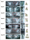
Frequency of impacted teeth and categorization of impacted canines: A retrospective radiographic study using orthopantomograms
- PMID: 28435377
- PMCID: PMC5379823
- DOI: 10.4103/ejd.ejd_308_16
Frequency of impacted teeth and categorization of impacted canines: A retrospective radiographic study using orthopantomograms
Abstract
Objective: The objective of this study is to determine the frequency of impacted maxillary canines using seven subtype classification system. For this purpose, impacted maxillary canines have been divided into seven various subtypes.
Materials and methods: This is a descriptive, cross-sectional, and retrospective study conducted using radiographic data of residents of Madinah, Al Munawwarah. Radiographic data of 14,000 patients, who attended College of Dentistry, Taibah University, from January 2011 to February 2015, were screened against the selection criteria for the presence of impacted teeth. The individuals with maxillary impacted canines were matched to maxillary canine impaction. The occurrence of each subtype of impacted canines was calculated.
Results: Impacted teeth are more common in the maxilla compared to mandible. The impacted canine represented the highest proportion of all impacted maxillary teeth followed by the second premolars and the central incisors. According to the classification system represented, Type II of canine impaction comprised the highest proportion (51%) while Type IV (0.5%) comprised the lowest frequency. The maxillary canine is the most frequently impacted tooth followed by mandibular canines.
Conclusions: Although there are many variations, the majority of impacted canines fall into Type II of the classification of impacted canines.
Keywords: Canine; impaction; maxillary teeth; orthodontics.
Conflict of interest statement
There are no conflicts of interest.
Figures


References
-
- Bass TB. Observations on the misplaced upper canine tooth. Dent Pract Dent Rec. 1967;18:25–33. - PubMed
-
- Pedro FL, Bandéca MC, Volpato LE, Marques AT, Borba AM, Musis CR, et al. Prevalence of impacted teeth in a Brazilian subpopulation. J Contemp Dent Pract. 2014;15:209–13. - PubMed
-
- Röhrer A. Displaced and impacted canines A radiographic research. Int J Orthod Oral Surg Radiogr. 1929;15:1003–20.
-
- Chung DD, Weisberg M, Pagala M. Incidence and effects of genetic factors on canine impaction in an isolated Jewish population. Am J Orthod Dentofacial Orthop. 2011;139:e331–5. - PubMed
-
- Sajnani AK, King NM. The sequential hypothesis of impaction of maxillary canine – A hypothesis based on clinical and radiographic findings. J Craniomaxillofac Surg. 2012;40:e375–85. - PubMed
LinkOut - more resources
Full Text Sources
Other Literature Sources

