

Review
doi: 10.5811/westjem.2016.12.31798.
Epub 2017 Mar 3.
Differentiating Urgent and Emergent Causes of Acute Red Eye for the Emergency Physician
Affiliations
- PMID: 28435504
- PMCID: PMC5391903
- DOI: 10.5811/westjem.2016.12.31798
Item in Clipboard
Review
Differentiating Urgent and Emergent Causes of Acute Red Eye for the Emergency Physician
West J Emerg Med.
2017 Apr.
Abstract
Patients commonly present with an acute red eye to the emergency department (ED). It is important to distinguish between benign and sight-threatening diagnoses. Here we provide a comprehensive overview on the acute red eye in the ED.
Conflict of interest statement
Conflicts of Interest: By the WestJEM article submission agreement, all authors are required to disclose all affiliations, funding sources and financial or management relationships that could be perceived as potential sources of bias. The authors disclosed none.
Figures
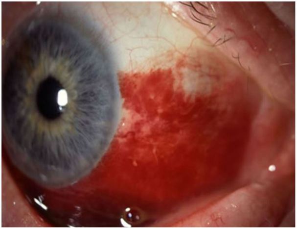
Subconjunctival hemorrhage. Image courtesy of Andrew Pearson, MA, MRCP.

Acute viral conjunctivitis. Image courtesy of Wikimedia Creative Commons.
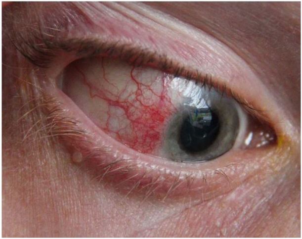
Episcleritis. Image courtesy of Asagan, Wikimedia Creative Commons.
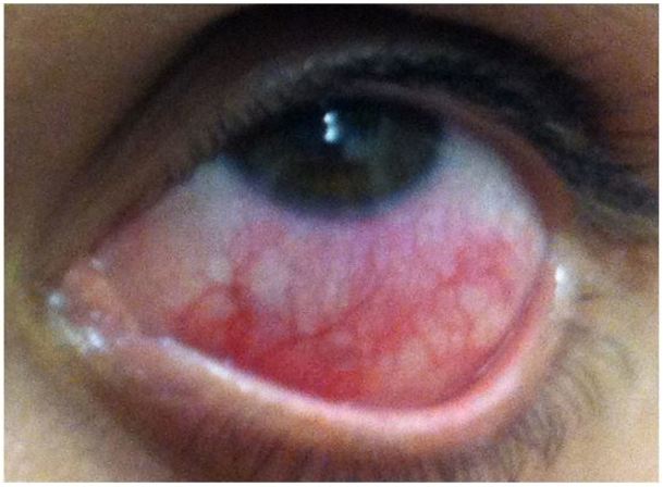
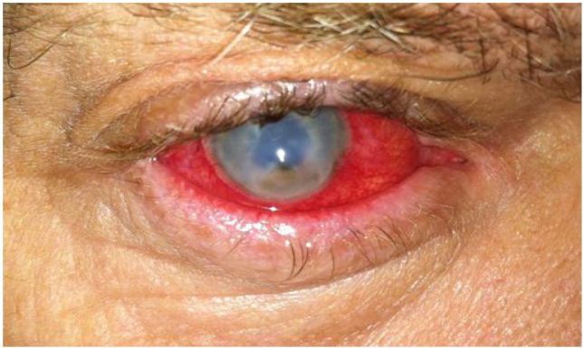
Anterior scleritis. Image courtesy of Marc Yonkers, MD, PhD.
Anterior uveitis. Image courtesy of Jonathan Trove, MD, Wikimedia Creative Commons.
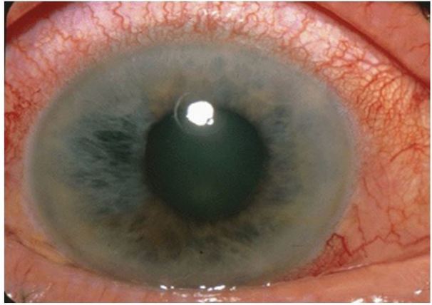
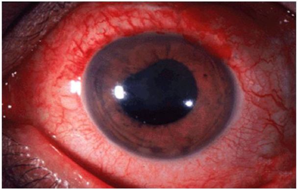
Acute angle closure glaucoma: note cloudy/“steamy” cornea and mid-position, fixed pupil. Image courtesy of Jonathan Trove, MD, Wikimedia Creative Common.
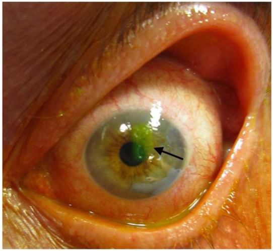
Corneal abrasion on fluorescein staining. Image courtesy of James Heilman, MD, Wikimedia Creative Commons.
Corneal ulcer. Image courtesy of Andrew Pearson, MA, MRCP.
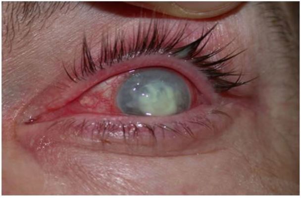
Endophthalmitis. Image courtesy of Marc Yonkers, MD, PhD.
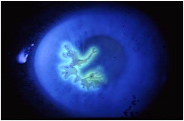
Viral keratitis (HSV keratitis). Image courtesy of Andrew Pearson, MA, MRCP.
References
-
- Alfonso SA, Fawley JD, Alexa Lu X. Conjunctivitis. Prim Care. 2015;42(3):325–45. - PubMed
-
- Diaz JD, Sobol EK, Gritz DC. Treatment and Management of Scleral Disorders. Surv Ophthalmol. 2016;61(6):702–17. - PubMed
-
- Ahmed F, House RJ, Feldman BH. Corneal abrasions and corneal foreign bodies. Prim Care. 2015;42(3):363–75. - PubMed
-
- Kongau Y, Henkind P. Pain elicited by consensual pupillary reflex: A diagnostic test for acute iritis. Lancet. 1981;2(8258):1254–5. - PubMed
-
- Prum BE, Jr, Herndon LW, Jr, Moroi SE, et al. Primary angle closure Preferred Practice Pattern(®) guidelines. Ophthalmology. 2016;123(1):1–40. - PubMed
Publication types
MeSH terms
LinkOut - more resources
Full Text Sources
Other Literature Sources
Medical
Molecular Biology Databases