Docosahexaenoic acid (DHA) promotes immunogenic apoptosis in human multiple myeloma cells, induces autophagy and inhibits STAT3 in both tumor and dendritic cells
- PMID: 28435516
- PMCID: PMC5396621
- DOI: 10.18632/genesandcancer.131
Docosahexaenoic acid (DHA) promotes immunogenic apoptosis in human multiple myeloma cells, induces autophagy and inhibits STAT3 in both tumor and dendritic cells
Abstract
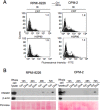
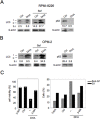
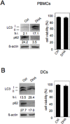
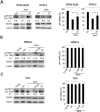
Docosahexaenoic acid (DHA), a ω-3 polyunsaturated fatty acid found in fish oil, is a multi-target agent and exerts anti-inflammatory and anticancer activities alone or in combination with chemotherapies. Combinatorial anticancer therapies, which induce immunogenic apoptosis, autophagy and STAT3 inhibition have been proposed for long-term therapeutic success. Here, we found that DHA promoted immunogenic apoptosis in multiple myeloma (MM) cells, with no toxicity on PBMCs and DCs. Immunogenic apoptosis was shown by the emission of specific DAMPs (CRT, HSP90, HMGB1) by apoptotic MM cells and the activation of their pro-apoptotic autophagy. Moreover, immunogenic apoptosis was directly shown by the activation of DCs by DHA-induced apoptotic MM cells. Furthermore, we provided the first evidence that DHA activated autophagy in PBMCs and DCs, thus potentially acting as immune stimulator and enhancing processing and presentation of tumor antigens by DCs. Finally, we found that DHA inhibited STAT3 in MM cells. STAT3 pathway, essential for MM survival, contributed to cancer cell apoptosis by DHA. We also found that DHA inhibited STAT3 in blood immune cells and counteracted STAT3 activation by tumor cell-released factors in PBMCs and DCs, suggesting the potential enhancement of the anti-tumor function of multiple immune cells and, in particular, that of DCs.
Keywords: STAT3; autophagy; dendritic cells-DCs; docosahexaenoic acid-DHA; immunogenic cell death.
Conflict of interest statement
CONFLICTS OF INTEREST No conflicts of interest were disclosed.
Figures


Similar articles
-
Capsaicin triggers immunogenic PEL cell death, stimulates DCs and reverts PEL-induced immune suppression.Oncotarget. 2015 Oct 6;6(30):29543-54. doi: 10.18632/oncotarget.4911. Oncotarget. 2015. PMID: 26338963 Free PMC article.
-
The n3-polyunsaturated fatty acid docosahexaenoic acid induces immunogenic cell death in human cancer cell lines via pre-apoptotic calreticulin exposure.Cancer Immunol Immunother. 2011 Oct;60(10):1503-7. doi: 10.1007/s00262-011-1074-7. Epub 2011 Jul 22. Cancer Immunol Immunother. 2011. PMID: 21779875 Free PMC article.
-
Human dendritic cell activities are modulated by the omega-3 fatty acid, docosahexaenoic acid, mainly through PPAR(gamma):RXR heterodimers: comparison with other polyunsaturated fatty acids.J Leukoc Biol. 2008 Oct;84(4):1172-82. doi: 10.1189/jlb.1007688. Epub 2008 Jul 16. J Leukoc Biol. 2008. PMID: 18632990
-
Docosahexaenoic Acid Induces Oxidative DNA Damage and Apoptosis, and Enhances the Chemosensitivity of Cancer Cells.Int J Mol Sci. 2016 Aug 3;17(8):1257. doi: 10.3390/ijms17081257. Int J Mol Sci. 2016. PMID: 27527148 Free PMC article. Review.
-
Tumor cell lysates as immunogenic sources for cancer vaccine design.Hum Vaccin Immunother. 2014;10(11):3261-9. doi: 10.4161/21645515.2014.982996. Hum Vaccin Immunother. 2014. PMID: 25625929 Free PMC article. Review.
Cited by
-
Docosahexaenoic Acid Inhibits Cytokine Expression by Reducing Reactive Oxygen Species in Pancreatic Stellate Cells.J Cancer Prev. 2021 Sep 30;26(3):195-206. doi: 10.15430/JCP.2021.26.3.195. J Cancer Prev. 2021. PMID: 34703822 Free PMC article.
-
The disruption of protein-protein interactions with co-chaperones and client substrates as a strategy towards Hsp90 inhibition.Acta Pharm Sin B. 2021 Jun;11(6):1446-1468. doi: 10.1016/j.apsb.2020.11.015. Epub 2020 Nov 24. Acta Pharm Sin B. 2021. PMID: 34221862 Free PMC article. Review.
-
Omega‑3 polyunsaturated fatty acids inhibit IL‑11/STAT3 signaling in hepatocytes during acetaminophen hepatotoxicity.Int J Mol Med. 2021 Oct;48(4):190. doi: 10.3892/ijmm.2021.5023. Epub 2021 Aug 20. Int J Mol Med. 2021. PMID: 34414450 Free PMC article.
-
Genetic evidence reveals phosphatidylcholine as a mediator in the causal relationship between omega-3 and multiple myeloma risk.Sci Rep. 2025 Aug 8;15(1):29016. doi: 10.1038/s41598-025-12804-y. Sci Rep. 2025. PMID: 40781111 Free PMC article.
-
Stimulators of immunogenic cell death for cancer therapy: focusing on natural compounds.Cancer Cell Int. 2023 Sep 13;23(1):200. doi: 10.1186/s12935-023-03058-7. Cancer Cell Int. 2023. PMID: 37705051 Free PMC article. Review.
References
-
- Laviano A, Rianda S, Molfino A, Rossi Fanelli F. Omega-3 fatty acids in cancer. Curr Opin Clin Nutr Metab Care. 2013;16:156–161. - PubMed
LinkOut - more resources
Full Text Sources
Other Literature Sources
Research Materials
Miscellaneous

