Genetically transforming human osteoblasts to sarcoma: development of an osteosarcoma model
- PMID: 28435520
- PMCID: PMC5396624
- DOI: 10.18632/genesandcancer.133
Genetically transforming human osteoblasts to sarcoma: development of an osteosarcoma model
Abstract
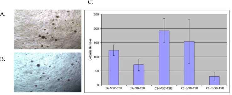
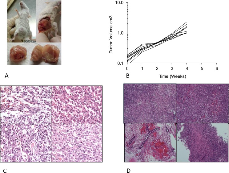
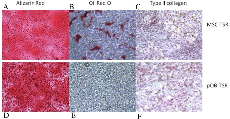
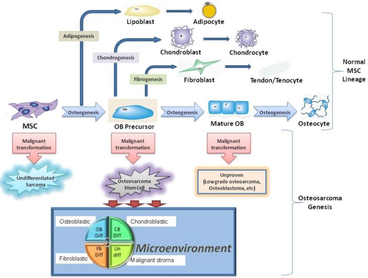
Osteosarcoma is the most common primary malignant bone tumor in children and young adults. Although histologically defined by the presence of malignant osteoid, the tumor possesses lineage multipotency suggesting it could be derived from a cell anywhere on the differentiation pathway between a mesenchymal stem cell (MSC) and a mature osteoblast. To determine if preosteoblasts (pOB) could be the cell of origin differentiated MSCs were transformed with defined genetic elements. MSCs and pOB differentiated from the same MSCs were serially transformed with the oncogenes hTERT, SV40 large T antigen and H-Ras. Assays were performed to determine their tumorigenic properties, differentiation capacity and histologic appearance. When subcutaneously implanted in immunocompromised mice, cell lines derived from transformed MSC and pOB formed tumors in 4 weeks. In contrast to the transformed MSC, the pOB tumors demonstrated a histological appearance characteristic of osteosarcoma. The cell lines derived from the transformed pOB only had osteogenic and chondrogenic differentiation potential, but not adipogenic ones. However, the transformed MSC cells and standard osteosarcoma cell lines maintained their tri-lineage differentiation capacity. The inability of the transformed pOB cell line to undergo adipogenic differentiation, may suggest that osteosarcoma is derived from a cell intermediate in differentiation between an MSC and a pOB, with partial commitment to the osteoblastic lineage.
Keywords: mesenchymal stem cells; osteoblast; osteosarcoma.
Conflict of interest statement
CONFLICTS OF INTEREST The authors confirm that there are no conflicts of interest.
Figures
References
-
- Geller DS, Gorlick R. Osteosarcoma: a review of diagnosis, management, and treatment strategies. Clinical advances in hematology % oncology: H&O. 2010;8:705. - PubMed
-
- Fletcher CDM UK, Mertens F. WHO Classification of Tumors. Lyon: IARC Press; 2002. Pathology and genetics of tumors of soft tissue and bone; pp. 1–427.
-
- Gorlick R BS, Teot L, Meyer J, Randall L, Marina N. Osteosarcoma: Biology, Diagnosis, Treatment and Remaining Challenges. In: Pizzo P, Poplack D, editors. Principles and Practice of Pediatric Oncology. Philadelphia: Lippincott Williams & Wilkins; 2011. pp. 1015–44.
-
- Gill J, Ahluwalia MK, Geller D, Gorlick R. New targets and approaches in osteosarcoma. Pharmacology and Therapeutics. 2012;1:89–99. - PubMed
LinkOut - more resources
Full Text Sources
Other Literature Sources
Research Materials
Miscellaneous