Case Reports
. 2017 Mar 29;9(1):6811.
doi: 10.4081/rt.2017.6811. eCollection 2017 Mar 24.

Angiofibrolipoma of Posterior Mediastinum with Transforaminal Extension

Affiliations
Case Reports

Angiofibrolipoma of Posterior Mediastinum with Transforaminal Extension

Monia Attia et al. Rare Tumors. .

Abstract

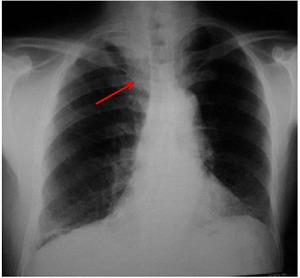
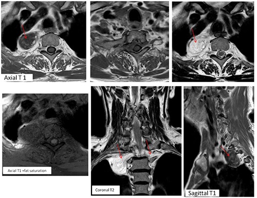
Angiofibrolipoma is a histological variant of lipoma, which commonly occurs in subcutaneous tissues. In the present report we illustrate the case of an angiofibrolipoma of the posterior upper mediastinum in a 75-year-old man presented with progressive chest pain. Xray chest showed a homogeneous opacity vertically oriented along the right lateral aspect of thoracic vertebrae with an obtuse angle to the mediastinum. The upper extremity of the mass extended above the superior clavicle, suggestive of a posterior mediastinal lesion. Thoracic magnetic resonance imaging revealed a posterior mediastinal mass, in keeping with a nonaggressive lesion, with particular endocanalar extension and heterogeneous signal and enhancement patterns that was highly suggestive of a mixed mesenchymal tumor. The tumor was incompletely removed by right postero-lateral thoracotomy with final diagnosis of angiofibrolipoma. To the author's knowledge, such a case of angiofibrolipoma located in the posterior mediastinum has not been previously reported in the literature.

Keywords: Angiofibrolipoma; MRI; Mesenchymal tumors; Posterior mediastinum.


Figures

Figure 1.
Figure 1.
Posteroanterior X-ray chest (frontal incidence): i) homogeneous opacity without air bronchogram, vertically oriented along the right lateral aspect of thoracic vertebrae with an obtuse angle to the mediastinum suggestive of a mediastinal opacity; ii) the upper extremity of the mass extends above the superior clavicle, therefore, it is probably located within the posterior mediastinum (cervicothoracic sign); iii) no mediastal shift; iv) no spinal or costal abnormalities seen.
Figure 2.
Figure 2.
Non-enhanced magnetic resonance imaging T1 and T2-weighted images before and after fat saturation: i) a mass located in the right costovertebral gutter, well-marginated, with heterogeneous low-signal intensity on T1- weighted images and a high-signal intensity on T2-weighted images. ii) Presence of hyperintense areas within the mass on both T1 and T2 WI that became hypointense after fat suppression corresponding to a fatty component. iii) The tumor extends through the spinal canal, filling the anterior epidural space, from right to left T1-T2 intervertebral foramina. iv) The spinal cord is slightly compressed without signal abnormalities. v) Scalloping of the posterior margin of the body and posterior arches of T1 and T2 VT. vi) The tumor is responsible of widening of both foramina and scalloping of posterior vertebral body and arches of T1 and T2 without cortical destruction.
Figure 3.
Figure 3.
Axial and sagittal fat-suppressed post contrast magnetic resonance imaging images: i) the mass shows an intense and heterogeneous enhancement; ii) tumoral extension through the spinal canal, filling the anterior epidural space, from right to left T1-2 intervertebral foramina that are enlarged; iii) there is no enhancement abnormalities of spinal cord next to the tumor.
Figure 4.
Figure 4.
Pathological image of the tumor (Hematoxylin and Eosin, 10×): i) association of the three characteristic findings of angiofibrolipomas: mature adipocytes, vascular vessels and collagenous tissue; ii) no evidence of mitosis or cellular pleomorphism.

Similar articles

References

    1. Duffy S, Jhaveri M, Scudierre J, et al. MR imaging of a posterior mediastinal ganglioneuroma: fat as a useful diagnostic sign. Am J Neuroradiol 2005;26:2658-62. - PMC - PubMed
    1. Nakazono T, White CS, Yamasaki F, et al. MRI findings of mediastinal neurogenic tumors. Am J Roentgenol 2011;197:W643-52. - PubMed
    1. Del Campo C, Mpougas PP. Compression of the superior vena cava by a mediastinal lipoma. Tex Heart Inst J 2000;27:297-8. - PMC - PubMed
    1. Jacob A, Kneile J, Welling DB. Angiofibrolipoma of the ear canal. Laryngoscope 2005;115:1461-2. - PubMed
    1. Uwale Eyesan S, Christopher Ayeni S, Adesope Adesina S, et al. Angiofibrolipoma of the calf. Rare Tumors 2013;5:e48. - PMC - PubMed

Publication types

LinkOut - more resources