

The three Rs: Recruitment, Retention and Residence of leukocytes in the liver
- PMID: 28435674
- PMCID: PMC5384287
- DOI: 10.1038/cti.2016.84
The three Rs: Recruitment, Retention and Residence of leukocytes in the liver
Abstract
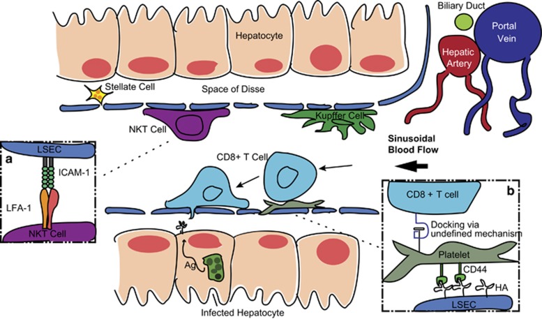
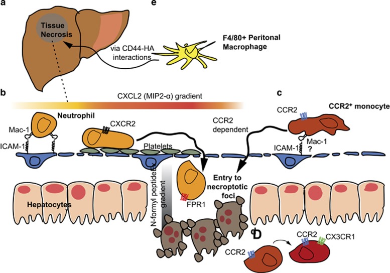
The composition of leukocytes in the liver is highly distinct from that of the blood and lymphoid organs. In particular, the liver is highly enriched in non-conventional T cells such as natural killer T (NKT) cells, γδ T cells and mucosal-associated invariant T cells. In addition, there are significant populations of tissue-resident NK cells (or innate lymphoid cells (ILC1)) and memory CD8+ T cells. These cells are joined in conditions of inflammation by neutrophils, monocytes and macrophages. In recent years a multitude of studies have generated insights into how these cells arrest, move and remain resident in the liver. This new understanding has largely been due to the use of intra-vital microscopy to track immune cells in the liver, coupled with gene expression profiling and parabiosis techniques. These studies have revealed that leukocyte recruitment in the liver does not correspond to the classical paradigm of the leukocyte adhesion cascade. Rather, both lymphoid and myeloid cells have been found to adhere in the liver sinusoids in a platelet-dependent manner. Leukocytes have also been observed to patrol the hepatic sinusoids using a characteristic crawling motility. Moreover, T cells have been observed surveying hepatocytes for antigen through the unique fenestrated endothelium of the liver sinusoids, potentially negating the need for extravasation. In this review we highlight some of these recent discoveries and examine the different molecular interactions required for the recruitment, retention and-in some cases-residence of diverse leukocyte populations within the liver.
Conflict of interest statement
The authors declare no conflict of interest.
Figures
References
-
- Sheth K, Bankey P. The liver as an immune organ. Curr Opin Crit Care 2001; 7: 99–104. - PubMed
-
- Jenne CN, Kubes P. Immune surveillance by the liver. Nat Immunol 2013; 14: 996–1006. - PubMed
-
- Warren A, Le Couteur DG, Fraser R, Bowen DG, McCaughan GW, Bertolino P. T lymphocytes interact with hepatocytes through fenestrations in murine liver sinusoidal endothelial cells. Hepatology 2006; 44: 1182–1190. - PubMed
Publication types
LinkOut - more resources
Full Text Sources
Other Literature Sources
Research Materials