. 2017 Mar 31;3(3):e1602552.
doi: 10.1126/sciadv.1602552. eCollection 2017 Mar.

Microbial arms race: Ballistic "nematocysts" in dinoflagellates represent a new extreme in organelle complexity

Affiliations

Microbial arms race: Ballistic "nematocysts" in dinoflagellates represent a new extreme in organelle complexity

Gregory S Gavelis et al. Sci Adv. .

Abstract

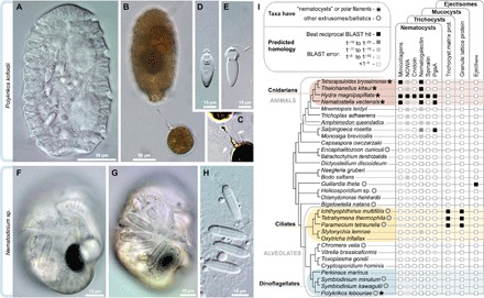
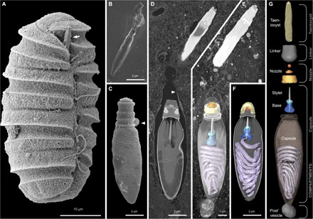
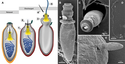
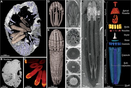
We examine the origin of harpoon-like secretory organelles (nematocysts) in dinoflagellate protists. These ballistic organelles have been hypothesized to be homologous to similarly complex structures in animals (cnidarians); but we show, using structural, functional, and phylogenomic data, that nematocysts evolved independently in both lineages. We also recorded the first high-resolution videos of nematocyst discharge in dinoflagellates. Unexpectedly, our data suggest that different types of dinoflagellate nematocysts use two fundamentally different types of ballistic mechanisms: one type relies on a single pressurized capsule for propulsion, whereas the other type launches 11 to 15 projectiles from an arrangement similar to a Gatling gun. Despite their radical structural differences, these nematocysts share a single origin within dinoflagellates and both potentially use a contraction-based mechanism to generate ballistic force. The diversity of traits in dinoflagellate nematocysts demonstrates a stepwise route by which simple secretory structures diversified to yield elaborate subcellular weaponry.

Keywords: Convergent evolution; cnidocyst; extrusome; minicollagen; mucocyst; red queen; secretion; secretory; trichocyst.


Figures

Fig. 1
Fig. 1. Diversity and independent origins of extrusomes.
(A to E) Nematocysts in the dinoflagellate P. kofoidii, including a live whole cell (A) and a cell that was preserved in Lugol’s iodide solution while capturing a prey cell of A. tamarense (B). (C) Enhanced contrast image shows the defensive trichocysts deployed by A. tamarense (arrows) in response to attack by P. kofoidii. (D and E) Isolated nematocysts from P. kofoidii, seen as unfired (D) and discharged (E). (F to H) Nematocysts in the dinoflagellate Nematodinium sp., which have an eye-like ocelloid (F). A battery of nematocysts is visible in the live cell (G) and remains intact after cell lysis (H). (I) Genomic distribution of known extrusome proteins (vertical labels) across eukaryotes based on best reciprocal Basic Local Alignment Search Tool (BLAST) hits (black squares), which is a common predictor of protein homology. Taxa are listed within an established phylogenetic framework (38, 39).
Fig. 2
Fig. 2. Reconstruction of the nematocysts in the dinoflagellate P. kofoidii.
(A) SEM micrograph of a cell of P. kofoidii, including an armed taeniocyst (arrow) in the apical region that first contacts prey. (B) SEM micrograph of an isolated taeniocyst that has discharged its amorphous contents. (C) SEM micrograph of an isolated nematocyst that has become arrested very early in discharge; arrowhead, operculum. (D) FIB-SEM section of taeniocyst and nematocyst enclosed by a membranous chute (arrowhead) and (E) maximum intensity projection of the same region seen slightly from above and below (F). (G) Virtual dissection of the nematocyst-taeniocyst complex. Brackets indicate the membrane-bound compartments in which those components are grouped during early development. Later in development, compartments fuse to form the chute.
Fig. 3
Fig. 3. Nematocyst discharge in Polykrikos.
(A) The putative ballistic sequence of nematocyst discharge is as follows: (1) striated capsule contracts; (2) internal pressure forces stylet to pierce capsule and open operculum (dark green); (3) part of the capsule everts; (4) stylet exits through the nozzle and detaches; and (5) mucilaginous tubule (blue) uncoils and is projected through the stylet base (yellow). Orange, relaxed capsule wall; red, contracted capsule wall. (B to E) SEM micrographs of isolated discharged nematocysts (B to D) and a nematocyst piercing a prey cell (E). Arrowhead, operculum; C, capsule; T, tubule.
Fig. 4
Fig. 4. Reconstruction of the nematocysts in Nematodinium sp.
(A to E) Images derived from single-cell FIB-SEM. (A) Partial reconstruction of a cell with nematocysts shown in beige and mucocysts shown in purple. (B) Longitudinal FIB-SEM section through a cell showing the nucleus and nematocysts (arrowheads). (C) 3D maximum intensity projection showing a battery of nematocysts. (D) 3D tilted reconstruction of a nematocyst. (E) Longitudinal reconstruction of a nematocyst. (F to K) Single-cell TEM micrographs of a nematocyst in oblique section (F), cross section (G to J), and longitudinal section (K) showing the striated material (arrow) and variable symmetries across different nematocysts, with the number of subcapsules printed in the upper right corner. (L) A virtual dissection of the nematocyst components. Brackets indicate the membrane-bound compartments bounding the components indicated. (F) to (J) scale bar, 500 nm.

Similar articles

Cited by

References

    1. Hausmann K., Extrusive organelles in protists. Int. Rev. Cytol. 52, 197–276 (1978). - PubMed
    1. Schmoker C., Hernández-León S., Calbet A., Microzooplankton grazing in the oceans: Impacts, data variability, knowledge gaps and future directions. J. Plankton Res. 35, 691–706 (2013).
    1. Matsuoka K., Cho H.-J., Jacobson D. M., Observations of the feeding behavior and growth rates of the heterotrophic dinoflagellate Polykrikos kofoidii (Polykrikaceae, Dinophyceae). Phys. Chem. Chem. Phys. 39, 82–86 (2000).
    1. Lee M. J., Jeong H. J., Lee K. H., Jang S. H., Kim J. H., Kim K. Y., Mixotrophy in the nematocyst–taeniocyst complex-bearing phototrophic dinoflagellate Polykrikos hartmannii. Harmful Algae 49, 124–134 (2015).
    1. Westfall J. A., Bradbury P. C., The fine structure of the nematocyst taeniocyst complex in polykrikos-kofoidi. J. Protozool. 29, 474–475 (1982).