Label-free optical detection of single enzyme-reactant reactions and associated conformational changes
- PMID: 28435868
- PMCID: PMC5371424
- DOI: 10.1126/sciadv.1603044
Label-free optical detection of single enzyme-reactant reactions and associated conformational changes
Abstract
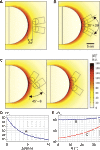
Monitoring the kinetics and conformational dynamics of single enzymes is crucial to better understand their biological functions because these motions and structural dynamics are usually unsynchronized among the molecules. However, detecting the enzyme-reactant interactions and associated conformational changes of the enzyme on a single-molecule basis remains as a challenge to established optical techniques because of the commonly required labeling of the reactants or the enzyme itself. The labeling process is usually nontrivial, and the labels themselves might skew the physical properties of the enzyme. We demonstrate an optical, label-free method capable of observing enzymatic interactions and associated conformational changes on a single-molecule level. We monitor polymerase/DNA interactions via the strong near-field enhancement provided by plasmonic nanorods resonantly coupled to whispering gallery modes in microcavities. Specifically, we use two different recognition schemes: one in which the kinetics of polymerase/DNA interactions are probed in the vicinity of DNA-functionalized nanorods, and the other in which these interactions are probed via the magnitude of conformational changes in the polymerase molecules immobilized on nanorods. In both approaches, we find that low and high polymerase activities can be clearly discerned through their characteristic signal amplitude and signal length distributions. Furthermore, the thermodynamic study of the monitored interactions suggests the occurrence of DNA polymerization. This work constitutes a proof-of-concept study of enzymatic activities using plasmonically enhanced microcavities and establishes an alternative and label-free method capable of investigating structural changes in single molecules.
Keywords: conformational change; optical microcavity; polymerase; protein-DNA interactions; single enzyme dynamics; single molecule reaction; whispering Gallery mode.
Figures


Similar articles
-
Single Molecule Thermodynamic Penalties Applied to Enzymes by Whispering Gallery Mode Biosensors.Adv Sci (Weinh). 2024 Sep;11(35):e2403195. doi: 10.1002/advs.202403195. Epub 2024 Jul 12. Adv Sci (Weinh). 2024. PMID: 38995192 Free PMC article.
-
Single-molecule nucleic acid interactions monitored on a label-free microcavity biosensor platform.Nat Nanotechnol. 2014 Nov;9(11):933-9. doi: 10.1038/nnano.2014.180. Epub 2014 Aug 31. Nat Nanotechnol. 2014. PMID: 25173831
-
Whispering-Gallery Mode Optoplasmonic Microcavities: From Advanced Single-Molecule Sensors and Microlasers to Applications in Synthetic Biology.ACS Photonics. 2024 Feb 3;11(3):892-903. doi: 10.1021/acsphotonics.3c01570. eCollection 2024 Mar 20. ACS Photonics. 2024. PMID: 38523742 Free PMC article. Review.
-
Mismatch-induced conformational distortions in polymerase beta support an induced-fit mechanism for fidelity.Biochemistry. 2005 Oct 11;44(40):13328-41. doi: 10.1021/bi0507682. Biochemistry. 2005. PMID: 16201758
-
[The role of weak specific and nonspecific interactions in recognition and conversation by enzymes of long DNA].Mol Biol (Mosk). 2004 Sep-Oct;38(5):756-85. Mol Biol (Mosk). 2004. PMID: 15554181 Review. Russian.
Cited by
-
Label-Free Optical Resonator-Based Biosensors.Sensors (Basel). 2020 Oct 19;20(20):5901. doi: 10.3390/s20205901. Sensors (Basel). 2020. PMID: 33086566 Free PMC article. Review.
-
Ceruloplasmin in flatland: the relationship between enzyme catalytic activity and surface hydrophilicity.RSC Adv. 2022 Sep 6;12(39):25388-25396. doi: 10.1039/d2ra04159f. eCollection 2022 Sep 5. RSC Adv. 2022. PMID: 36199311 Free PMC article.
-
Thermo-Optoplasmonic Single-Molecule Sensing on Optical Microcavities.ACS Nano. 2024 Jul 9;18(27):17534-17546. doi: 10.1021/acsnano.4c00877. Epub 2024 Jun 26. ACS Nano. 2024. PMID: 38924515 Free PMC article.
-
Label-Free Plasmonic Detection of Untethered Nanometer-Sized Brownian Particles.ACS Nano. 2020 Oct 27;14(10):14212-14218. doi: 10.1021/acsnano.0c07335. Epub 2020 Oct 15. ACS Nano. 2020. PMID: 33054166 Free PMC article.
-
Roadmap on optical sensors.J Opt. 2017 Aug;19(8):083001. doi: 10.1088/2040-8986/aa7419. Epub 2017 Jul 24. J Opt. 2017. PMID: 29375751 Free PMC article.
References
-
- Farooq S., Fijen C., Hohlbein J., Studying DNA–protein interactions with single-molecule Förster resonance energy transfer. Protoplasma 251, 317–332 (2014). - PubMed
MeSH terms
Substances
LinkOut - more resources
Full Text Sources
Other Literature Sources

