

Bone bridge formation across the neuroforamen 14 years after instrumented fusion for isthmic spondylolisthesis-a case report
- PMID: 28435923
- PMCID: PMC5386897
- DOI: 10.21037/jss.2017.03.03
Bone bridge formation across the neuroforamen 14 years after instrumented fusion for isthmic spondylolisthesis-a case report
Abstract
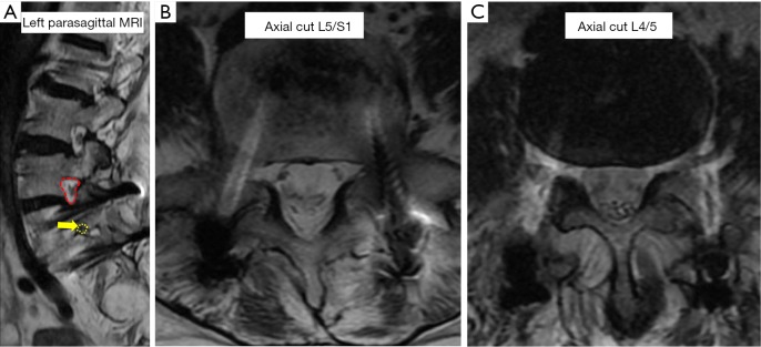
This case report describes the first case of a bone bridge formation across the left L5/S1 neuroforamen after instrumented posterolateral fusion for L5/S1 isthmic spondylolisthesis. Our patient was a 70-year-old lady who had grade 2, L5/S1 isthmic spondylolisthesis and bilateral S1 nerve root compression. She suffered from mechanical low back pain and neurogenic claudication, with radicular pain over both S1 dermatomes. She underwent in-situ, instrumented, posterolateral fusion and was asymptomatic for more than 13 years before developing progressive onset of left radicular pain over the L5 dermatome. Imaging revealed a bisected left L5/S1 neuroforamen secondary to a bone bridge formation resulting in stenosis. The pars defect in this case may have had sufficient osteogenic and osteoinductive factors to heal following spinal stabilization. Although in-situ posterolateral fusion is an accepted surgical treatment for isthmic spondylolisthesis, surgeons should consider reduction of the spondylolisthesis and excision of the pars defects to avoid this possible long-term complication.
Keywords: Spondylolysis; deformity; heterotopic ossification; spine; spondylolisthesis.
Conflict of interest statement
Conflicts of Interest: The authors have no conflicts of interest to declare.
Figures


Similar articles
-
Comparison of instrumented posterolateral fusion versus percutaneous pedicle screw fixation combined with anterior lumbar interbody fusion in elderly patients with L5-S1 isthmic spondylolisthesis and foraminal stenosis.J Neurosurg Spine. 2011 Sep;15(3):311-9. doi: 10.3171/2011.4.SPINE10653. Epub 2011 May 20. J Neurosurg Spine. 2011. PMID: 21599444
-
Acute progression of spondylolysis to isthmic spondylolisthesis in an adult.Spine (Phila Pa 1976). 2002 Aug 15;27(16):E370-2. doi: 10.1097/00007632-200208150-00023. Spine (Phila Pa 1976). 2002. PMID: 12195078 Review.
-
[Adjacent segment degeneration after lumbosacral fusion in spondylolisthesis: a retrospective radiological and clinical analysis].Acta Chir Orthop Traumatol Cech. 2010 Apr;77(2):124-30. Acta Chir Orthop Traumatol Cech. 2010. PMID: 20447355 Czech.
-
High-grade adult isthmic L5-s1 spondylolisthesis: a report of intraoperative slip progression treated with surgical reduction and posterior instrumented fusion.Global Spine J. 2012 Jun;2(2):119-24. doi: 10.1055/s-0032-1307257. Global Spine J. 2012. PMID: 24353957 Free PMC article.
-
Spondylolysis and spondylolisthesis in children and adolescents: II. Surgical management.J Am Acad Orthop Surg. 2006 Aug;14(8):488-98. doi: 10.5435/00124635-200608000-00006. J Am Acad Orthop Surg. 2006. PMID: 16885480 Review.
References
Publication types
LinkOut - more resources
Full Text Sources
Other Literature Sources
Miscellaneous